Sensitive skin is a prevalent issue arising from factors such as environmental pollution, unhealthy lifestyles, and autoimmune responses, often linked to the activation of the TRPV1 pathway and subsequent immune-inflammatory reactions. Individuals experiencing sensitive skin frequently encounter discomfort and irritation, highlighting the need for effective soothing and anti-inflammatory treatments. This research considered the potential of utilizing β-chitooligosaccharides (COS) derived from marine Sargassum due to their known ability to regulate inflammatory responses and interact with TRPV1 pathways. Furthermore, the incorporation of dopamine, a naturally occurring catecholamine with adhesive properties and presence in Sargassum extracts, was explored to enhance the therapeutic potential and skin compatibility of the resulting compound. The study aimed to synthesize and evaluate the properties and anti-inflammatory effects of COS-diDA, a novel compound formed by binding two dopamine amines to COS, as a potential treatment for sensitive skin.
Methods
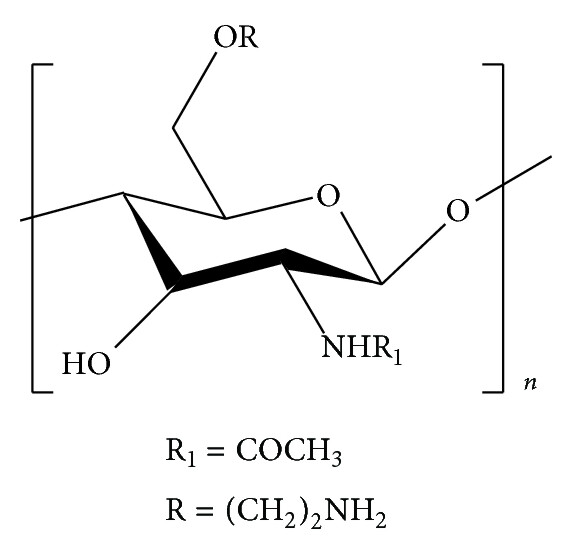
The synthesis of COS-diDA involved Michael addition and Schiff base reactions between COS and dopamine under alkaline conditions. The resulting compound was characterized using UV-Vis spectroscopy, ATR-FTIR, high-resolution mass spectrometry, and scanning electron microscopy (SEM) to confirm its structure and morphology. In vitro studies were conducted using a HaCaT cell model stimulated with capsaicin (CAP) to mimic sensitive skin conditions. Cell proliferation was assessed using the CCK-8 assay, while ELISA and immunofluorescence experiments were employed to determine the impact of COS-diDA on TRPV1 protein levels and the secretion of inflammatory cytokines IL-1α, IL-6, and IL-8. Statistical analysis was performed using GraphPad Prism 9.0 to evaluate the significance of the findings.
Section-wise Key Points
•Characterization of COS-diDA Properties: Spectroscopic analyses (UV-Vis and FTIR) confirmed the presence of catechol groups from dopamine in the synthesized COS-diDA. High-resolution mass spectrometry indicated that each monosaccharide unit of COS was connected to one dopamine molecule, suggesting two dopamine molecules bind to every five sugar units. SEM revealed that COS-diDA exhibited a smooth, dense, and regular layered structure with reduced porosity compared to COS, suggesting enhanced adhesion properties.
•Effect of COS-diDA on the Proliferation and Activity of HaCaT Cells: COS-diDA showed no toxic effects on HaCaT cells and enhanced cell proliferation at concentrations up to 1.6 μg/mL.
•Determination of CAP Concentration: A CAP concentration of 125 μg/mL was determined to be the half-maximal inhibitory concentration (IC50) for HaCaT cell proliferation and was used to establish the in vitro sensitive skin model.
•Effect of COS-diDA on CAP-Induced Growth Inhibition of HaCaT Cells: Treatment with COS-diDA significantly increased the viability of CAP-stimulated HaCaT cells across various concentrations, demonstrating its ability to mitigate CAP-induced damage. Notably, 0.8 μg/mL of COS-diDA showed significant therapeutic effects, even outperforming the positive control, TTBC.
•Inhibitory Effect of COS-diDA on TRPV1 Protein in CAP-Activated HaCaT Cells: CAP stimulation significantly increased TRPV1 protein levels, validating the pain model. COS-diDA treatment led to a reduction in TRPV1 protein levels in CAP-stimulated HaCaT cells in a concentration-dependent manner, indicating its soothing effect by suppressing TRPV1 activation.
•Effect of COS-diDA on the Level of Interleukin Secretion by CAP-Induced HaCaT Cells: CAP exposure significantly elevated the secretion of pro-inflammatory cytokines IL-1α, IL-6, and IL-8 from HaCaT cells. COS-diDA treatment significantly suppressed the release of these inflammatory mediators, indicating its potent anti-inflammatory action in the CAP-induced sensitive skin model. The optimal concentration for inhibiting these interleukins varied slightly, with 0.8 μg/mL showing prominent effects for IL-1α and IL-6 at 24 hours, and varying concentrations showing efficacy for IL-8.
This research demonstrates the successful synthesis and characterization of marine-derived COS-diDA, a novel conjugate of β-chitooligosaccharides and dopamine. The study’s novelty lies in its exploration of this specific marine-derived compound for sensitive skin treatment and the comprehensive evaluation of its anti-inflammatory and soothing effects at a cellular level. The findings reveal that COS-diDA effectively mitigates CAP-induced cellular damage, suppresses the expression of the TRPV1 receptor, and inhibits the secretion of key pro-inflammatory cytokines (IL-1α, IL-6, and IL-8) in keratinocytes. These results strongly suggest the potential of COS-diDA as a promising anti-inflammatory and pain-relieving agent for sensitive skin. Future implications of this research include further in vivo studies to validate these effects and explore the formulation of COS-diDA as a valuable supplementary component in cosmetic products aimed at alleviating sensitive skin conditions.
Link to the study: https://www.mdpi.com/2079-9284/12/2/35