Wound healing is a complex and highly coordinated biological process that proceeds through distinct phases: homeostasis, inflammation, proliferation, and remodeling. Each phase is critically regulated by an intricate network of signaling pathways, and any disruption to this delicate balance can lead to chronic, non-healing wounds. Therefore, understanding and modulating these pathways are essential for developing effective therapeutic strategies to enhance wound repair.
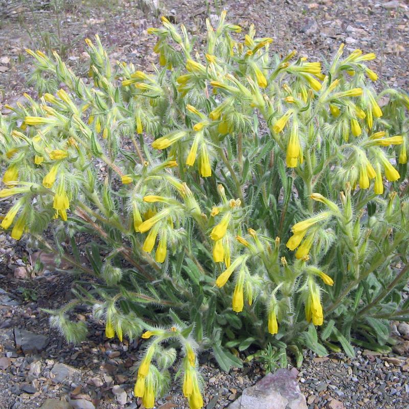
In the search for such solutions, traditional medicine offers a rich source of potential agents. The root extract of Onosma microcarpum (OM), a plant traditionally used in Western Iran for treating infectious wounds, inflammation, and episiotomy pain, has garnered attention for its wound-healing properties. OM is known to contain a variety of bioactive compounds, including alkannin, shikonin, polyphenols, alkaloids, and terpenoids, which possess strong antioxidant, anti-inflammatory, antimicrobial, and pro-regenerative effects. While previous studies indicated promising wound-healing effects of OM, particularly in diabetic rat models, the specific mechanisms and underlying signaling pathways involved remained largely unexplored. This research aimed to elucidate how OM extract enhances wound healing by modulating key upstream pathways, specifically the Mitogen-Activated Protein Kinase (MAPK), Janus Kinase/Signal Transducer and Activator of Transcription (JAK/STAT), and Phosphoinositide 3-Kinases (PI3K)/Akt pathways, which are critical regulators of the healing process.
Methods
The study involved male Wistar rats with excisional wounds, treated topically with either 0.2% or 1% Onosma microcarpum (OM) ointment or a vehicle control. Wound closure rates, histological parameters (inflammation, granulation tissue formation, angiogenesis, and collagen formation), and gene expression levels of key cytokines, growth factors, and cell regulators (including IL-1β, TNF-α, IL-6, TGF-β1, PDGF, ERK, JNK, and p38 MAPK) were assessed on days 3, 8, and 14 post-wounding. Plant roots were collected, extracted, and formulated into ointments under sterile conditions. Statistical analysis was conducted using SPSS, employing ANOVA or Kruskal-Wallis tests, with a P-value of less than 0.05 considered significant.
Key Findings
The research yielded significant dose-specific insights into the therapeutic potential of Onosma microcarpum extract in wound healing:
• Optimal Dose and Overall Efficacy: The 0.2% OM concentration consistently demonstrated superior wound-healing outcomes compared to both the vehicle control and the 1% OM group. Wounds treated with 0.2% OM showed a higher wound closure rate (87% vs. 69% in vehicle, P=0.023), more organized collagen (mean score 3.0 vs. 2.3, P=0.021), and improved angiogenesis (3.4 vs. 2.5, P=0.038) by day 14.
• Modulation During the Inflammatory Phase (Day 3):
◦ 0.2% OM significantly reduced inflammation scores and the expression of pro-inflammatory cytokines, TNF-α (P=0.003) and IL-1β (P=0.008), while also decreasing JNK activity (P=0.009). This indicates a beneficial anti-inflammatory effect early in the healing process.
◦ In contrast, the 1% OM group increased p38 MAPK (P=0.038) and collagen type I (P=0.022) expression compared to the vehicle group, which might promote scarring rather than efficient wound healing if activated too early.
• Modulation During the Proliferative Phase (Day 8):
◦ 0.2% OM further suppressed inflammation by reducing IL-1β (P=0.001) and IL-6 (P=0.002) expression, and decreased p38 MAPK (P=0.005) and MMP9 (P=0.003) levels.
◦ Crucially, 0.2% OM significantly elevated the expression of growth factors TGF-β1 (P=0.008) and PDGF (P=0.001), promoting fibroblast proliferation, granulation tissue formation (P=0.038), and angiogenesis (P=0.023).
◦ The 1% OM group did not show significant improvements in granulation or angiogenesis and was associated with a reduction in AKT and TGF-β expression, suggesting potential detrimental effects at higher concentrations.
• Modulation During the Regenerative Phase (Day 14):
◦ 0.2% OM increased ERK (P=0.008) and p38 MAPK (P=0.002) activity, along with continued elevation of TGF-β1 (P=0.001) and PDGF (P=0.003), and a surge in α-SMA (P<0.001) and collagen type I (P=0.007) expression, facilitating organized tissue repair and regeneration.
◦ The 1% OM group resulted in moderately increased but disorganized collagen structure, failing to demonstrate significant improvements in collagen formation or angiogenesis compared to the vehicle.
• Limited Impact on Other Pathways: OM extract showed minimal impact on the JAK/STAT and AKT/PI3K pathways, as well as VEGF expression, suggesting a focused modulatory effect primarily on MAPK pathways.
The future implications of this research are substantial. Further studies should focus on identifying and quantifying the specific bioactive compounds within OM extract responsible for these effects to enable standardization and optimization for therapeutic use. Investigating the concentration-dependent biphasic effects of individual compounds or their synergistic interactions is crucial. Given OM’s minimal impact on JAK/STAT and AKT/PI3K pathways, exploring its combination with other therapeutic agents targeting these pathways could lead to more robust wound-healing outcomes. Finally, clinical trials are essential to validate the efficacy and safety of Onosma microcarpum extract in human wound healing, paving the way for its potential integration into clinical wound care practices.
Link to the study: https://tinyurl.com/dr4ekv45