Atopic dermatitis (AD) is a chronic inflammatory skin disease affecting a significant portion of children, accounting for 15.6–24% of childhood diseases. Its pathogenesis is complex, involving an interplay of skin barrier dysfunction and immune abnormalities, influenced by both genetic and environmental factors. The skin hosts a diverse community of microorganisms that are crucial for protecting the skin barrier and regulating the immune system. Recent research has increasingly highlighted the intimate connection between the skin microbiota and the onset and aggravation of AD. In patients with AD, this microbial balance is often disrupted, leading to an increased proportion of pathogenic bacteria, most notably Staphylococcus aureus (S. aureus). S. aureus colonization is prevalent in AD, occurring in 70% of lesioned skin and 39% of non-lesioned skin, and contributes to AD pathogenesis through its damage-response framework, exacerbating inflammation and damaging the skin barrier.
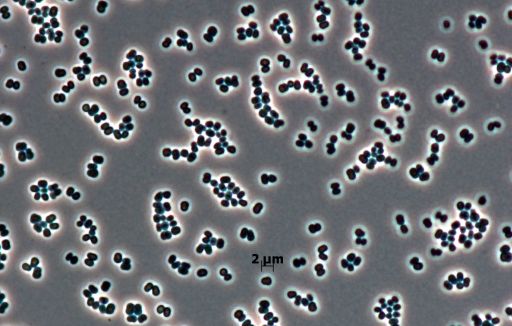
Given the critical role of skin microbiota in AD onset and progression, it has become an integral factor in exploring AD treatment approaches. Among the diverse skin microbiota, the genus Kocuria, belonging to the Micrococcaceae family, is a Gram-positive, catalase-positive, and coagulase-negative cocci. These ubiquitous bacteria are commonly found in the environment and are recognized as commensal bacteria of the normal skin microbiota. While most Kocuria species are symbiotic, some have industrial applications, with their carotenoid pigments used in food coloring and pharmaceuticals, and certain strains showing potential as probiotics or anti-cancer agents. This study represents the first attempt to specifically investigate the association between AD and Kocuria rhizophila, aiming to understand its relative abundance in the skin microbiota of children with and without AD, its correlation with AD severity, and its potential positive effects on skin barrier function against S. aureus virulence factors.
Methods
This prospective cross-sectional study involved 52 children (35 with AD and 17 without AD) aged 2–12 years, with AD severity quantified using SCORAD and EASI scores. Skin microbiome samples were analyzed using 16S rRNA amplicon sequencing. The anti-virulence effects of Kocuria rhizophila were estimated, and the effects of heat-killed K. rhizophila (HKKR) were further evaluated in human skin cell models (HaCaT cells) with AD-like damage induced by S. aureus secretory toxins (protein A, lipoteichoic acid, and protease V8). Additionally, a 3D human skin model was developed and treated with virulence factors, with histopathological and transcriptomic analyses performed.
Key Findings
• Microbial Diversity and Composition: Alpha-diversity indices tended to decrease in the AD-flare (AD-F) group compared with the non-AD group but were higher in the AD-non-flare (AD-NF) group. The AD group had a high relative abundance of S. aureus, which was almost absent in the non-AD group and significantly decreased in the AD-NF group.
• Kocuria rhizophila Correlation: K. rhizophila was negatively correlated with AD severity markers such as SCORAD, EASI, and Transepidermal Water Loss (TEWL), indicating that a higher abundance of K. rhizophila was associated with reduced AD aggravation and skin barrier dysfunction. It was highly abundant in non-AD skin (11.91% relative abundance).
• HKKR Effects on Skin Barrier Function: Treatment with heat-killed K. rhizophila (HKKR) upregulated the gene expression of the tight junction protein zonula occludens-1 (ZO-1) and critical components of the cornified cell envelope, involucrin (IVL) and filaggrin (FLG). HKKR restored the decrease in cell viability and transepithelial electrical resistance (TEER) caused by S. aureus protein A.
• HKKR Effects on Inflammation: HKKR treatment significantly down-regulated the expression of pro-inflammatory cytokines interleukin (IL)-1b and IL-6, which are typically elevated by S. aureus virulence factors. Transcriptomic analysis in the 3D human skin model revealed that the HKKR-treated group exhibited fewer alterations in genes associated with inflammatory regulation compared to the S. aureus virulence factor-treated group.
• Skin Protective Effects in 3D Model: In a 3D human skin model, HKKR mitigated V8-induced epidermal barrier disruption, restoring FLG expression to levels comparable to the control group. Transcriptomic data confirmed HKKR’s association with FLG expression and showed that HKKR significantly inhibited the reduction of FLG-associated protein gene expression, including CASP14, ST14, and TCHH, which are involved in skin barrier formation. HKKR also promoted gene sets associated with ‘keratinocyte differentiation,’ ‘keratinization,’ and ‘establishment of the skin barrier,’ counteracting V8-induced disruption.
This study pioneers in elucidating the association between AD and Kocuria rhizophila, marking the first research to explore the role of K. rhizophila in non-AD and AD with and without flares, and to demonstrate its positive effects on AD-affected skin. The novelty of this research lies in identifying K. rhizophila as a potential agent that can mitigate skin barrier dysfunction and inflammation, particularly in the context of S. aureus infection, through the upregulation of key skin barrier genes like ZO-1, IVL, and FLG, and the downregulation of pro-inflammatory cytokines IL-1b and IL-6. It also highlighted its role in promoting keratinocyte differentiation and keratinization.
The future implications of this research are significant, suggesting K. rhizophila as a promising novel therapeutic approach for AD treatment. While the precise mechanism by which K. rhizophila modulates FLG expression requires further investigation, its demonstrated ability to protect against skin barrier damage is a crucial finding. Furthermore, the study notes that K. rhizophila is a common soil bacterium, which aligns with the hygiene hypothesis, suggesting that natural environmental exposure to such microbes could offer protective benefits against AD onset and aggravation. Further studies are needed to explore K. rhizophila in the context of natural environmental exposure as a potential AD treatment, building upon these initial findings. Despite limitations such as a relatively small sample size, this study provides a foundational understanding of K. rhizophila‘s therapeutic potential in AD.
Link to the study: https://tinyurl.com/nknanv8p