Maintaining skin health, particularly through proper moisturization, is increasingly recognized as fundamental in the rapidly growing cosmetics industry. Consumers widely utilize skin moisturizers to preserve youthful, hydrated skin, prevent dryness, and reduce transepidermal water loss (TEWL). These products are crucial for upholding skin barrier integrity and mitigating the risk of conditions such as xerosis, inflammation, and wrinkle formation. The stratum corneum (SC), the outermost layer of the epidermis, is vital for skin hydration, and its disruption by external factors leads to increased TEWL and alterations in lipid and protein composition. Environmental stressors like surfactants, organic solvents, ultraviolet (UV) radiation, and mechanical factors can impair the SC’s lipid bilayers, leading to barrier dysfunction and dry skin conditions.
The development of effective moisturizing agents necessitates robust and reliable experimental models for efficacy evaluation. However, conventional in vitro cell-based assays face significant limitations due to the viscous and waxy nature of many cosmetic moisturizers, often failing to accurately replicate key functional endpoints such as TEWL and barrier recovery. Consequently, current evaluation primarily relies on clinical studies in human subjects, which are prone to inherent variability and demand stringent environmental control and large sample sizes. Recognizing these challenges and the ethical restrictions on animal testing in cosmetics, there is a pressing need for experimental models that can better simulate dry skin architecture, accommodate viscous formulations, and enable quantitative assessment of skin barrier function. The study by Choe et al. addresses this critical unmet need by proposing a novel in vitro dry skin model using isolated minipig epidermis and human cadaver skin as a practical, reproducible, and ethically compliant platform for evaluating moisturizing agents.
Methods
The study developed an in vitro dry skin model utilizing isolated minipig epidermis and human cadaver skin tissues. Various physical and chemical stressors, including lipid-extracting solvents (tert-butyl methyl ether, MTBE/Acetone solution), an irritant surfactant (sodium dodecyl sulfate), ultraviolet B (UVB) irradiation, and tape stripping, were applied to induce dry skin conditions. Skin barrier disruption and stratum corneum damage were then meticulously evaluated through assessments of epidermal lipid integrity using Nile Red staining and ImageJ analysis, histological alterations via H&E staining, transepidermal water loss (TEWL) measurements, and fluorescein isothiocyanate (FITC)-dextran permeation tests.
Key Findings
• All tested treatments, including lipid-extracting solvents, the irritant surfactant SDS, UVB irradiation, and tape stripping, successfully induced significant dry skin conditions, characterized by disrupted lipid architecture, histological damage, increased TEWL, and enhanced FITC-dextran flux.
• Among the various stimuli, the 50/50 MTBE/Acetone solution (M/A) applied for 5 minutes produced the most consistent and reproducible changes across all parameters in minipig epidermis. This protocol resulted in approximately a 50% reduction in the stratum corneum (SC) area with minimal inter-sample variability and consistently increased FITC-dextran permeability.
• The M/A 5-minute treatment also yielded comparable patterns of SC disruption in human cadaver skin, although human SC exhibited greater thickness and resistance to complete detachment, suggesting potential structural differences.
• Correlation analyses revealed a strong inverse correlation between SC area (quantified by Nile Red staining) and TEWL (r = -0.85, p = 0.004), as well as between SC area and FITC-dextran penetration (r = -0.81, p = 0.008), indicating that SC area serves as a reliable objective indicator of skin barrier damage.
• The model demonstrated the capability for dual assessment through both structural imaging and functional measurements, providing a comprehensive understanding of barrier impairment.
The future implications of this model are substantial. It represents a valuable tool to accelerate the development and screening of new skin moisturizers, potentially reducing the need for early-stage human testing. For practical implementation, future validation studies are necessary to test the applicability of this model with various reference substances of known moisturizing efficacy and to assess its intra- and inter-laboratory reproducibility. While the model currently cannot assess moisturizing-related genes, this limitation could be addressed by supplementing it with cell-based assays or ex vivo live skin models. Furthermore, extending the comparison to a wider range of concentrations and exposure times, including stronger damage conditions, will be crucial for optimizing the human tissue model and establishing a more robust basis for cross-species validation. This model, therefore, stands as a promising advancement in dermatological research and cosmetic product development.
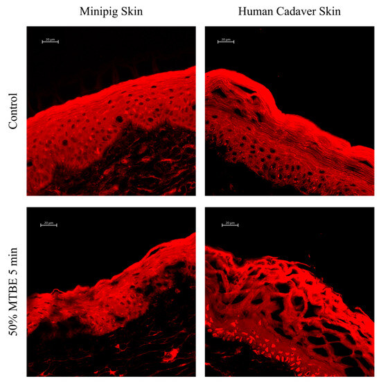
In the figure: Comparison of MTBE-induced dry skin model in minipig epidermis and human cadaver skin. M/A were applied to minipig epidermis or human cadaver skin for 5 min as skin drying condition. Treated skin tissues were sectioned to a thickness of less than 10 μm, mounted on glass slides, and stained with Nile red. Fluorescence images were captured at 40× magnification. The scale bar represents 20 µm.
Link to the study: https://www.mdpi.com/2079-9284/12/5/203