Fungal skin infections are a growing concern due to rising resistance and limited new antifungal agents. Candida spp., especially Candida albicans and the increasingly common C. glabrata, cause superficial infections like cutaneous candidiasis, which can lead to itching, inflammation, and pustules. C. glabrata is notable for its resistance to azoles and its virulence factors such as adhesion and biofilm formation. Dermatophytes like Trichophyton rubrum also infect keratinized tissues using keratin as a nutrient source. Treatment of these infections depends on immune response and antifungal therapy but is challenged by microbial resistance and pathogenicity.
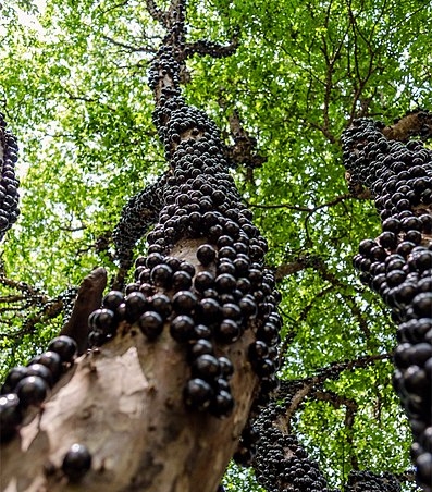
Natural sources like plants are being explored for new bioactive compounds, with Brazil’s biodiversity offering untapped potential. Plinia cauliflora (jabuticabeira), used traditionally for skin conditions, has shown in vitro antifungal activity, attributed to compounds like casuarinin, myricetin, and quercetin—flavonoids with known antimicrobial properties.
However, natural product stability remains a hurdle. Nanotechnology, particularly microemulsions—stable colloidal systems of oil and water stabilized by surfactants—can enhance solubility, protect active components, and boost skin absorption. This study investigates P. cauliflora leaf compounds and develops microemulsions incorporating them to enhance antifungal activity against skin infections.
Methods
Leaves of Plinia cauliflora were collected, dried, and crushed, followed by extraction using ethanol. The ethanolic extract (EtOH70) was fractionated using liquid-liquid partitioning with ethyl acetate, n-butanol, and water, yielding the FrAcOEt, FrBuOH, and FrAqu fractions. The extract and fractions were characterized by High-Performance Liquid Chromatography (HPLC). Microemulsions were developed using cholesterol, surfactants (Brij 58® and Epikuron® 200), and phosphate buffer, then sonicated and centrifuged. Samples (extract and fractions) were incorporated into the microemulsion at a final concentration and further sonicated. The microemulsions were characterized by particle diameter and polydispersity index (PDI) using dynamic light scattering. Antifungal activity against Candida glabrata and Trichophyton rubrum strains (including a clinical isolate) was evaluated by determining Minimum Inhibitory Concentration (MIC) and Minimum Fungicide Concentration (MFC) using broth microdilution techniques, with amphotericin B and fluconazole as controls. Antioxidant activity was assessed by the DPPH radical scavenging method, calculating the effective concentration (EC50). In vivo toxicity was evaluated using the Galleria mellonella larvae model.
Key Findings
• The Plinia cauliflora EtOH70 extract and its fractions showed antifungal activity against the tested strains of Candida glabrata and Trichophyton rubrum.
• C. glabrata was particularly sensitive to the samples.
• Incorporation of the EtOH70 extract into a microemulsion (mEtOH70) significantly decreased the MIC for C. glabrata, representing about a four-fold reduction compared to the non-incorporated extract.
• For the Trichophyton rubrum clinical isolate (T. rubrum 1), incorporation of the EtOH70 extract into a microemulsion (mEtOH70) reduced the MIC, a two-fold reduction.
• For the T. rubrum ATCC strain, mEtOH70 also showed the same MIC as the non-incorporated EtOH70 extract. Note: There appears to be a discrepancy or no improvement noted for T. rubrum ATCC MIC for EtOH70 vs mEtOH70 in the tables, although the abstract and conclusion state a reduction by half for T. rubrum with the incorporated samples. The discussion section clarifies the T. rubrum 1 result.
• The empty microemulsion (EM) showed high MIC values for both fungi, indicating the activity is primarily from the plant material.
• The incorporation of the butanolic fraction (mFrBuOH) into a microemulsion also showed enhanced activity, reducing the MIC for C. glabrata and for T. rubrum 1.
• The microemulsions, both empty and loaded with extract/fractions, showed small particle sizes and suitable polydispersity indices.
• All microemulsions exhibited negative zeta potential, suggesting effective electrostatic stabilization.
• The EtOH70 extract showed the best antioxidant activity among the plant samples tested in the DPPH assay.
• In the Galleria mellonella in vivo toxicity test, the extract, fractions, and incorporated microemulsions at the tested concentration showed no toxicity, with full larval survival and no observed melanization
This study presents findings suggesting that the leaves of Plinia cauliflora are a promising source of compounds for antifungal therapy. Notably, this research is the first to investigate the incorporation of P. cauliflora leaf extracts and fractions into microemulsions to enhance antifungal activity. The results demonstrated that this nanotechnology technique was effective in improving the antifungal activity of the extract and certain fractions, particularly against Candida glabrata, where the MIC was reduced significantly, and also showing enhanced efficacy against Trichophyton rubrum. The study also indicated some antioxidant activity of the samples and, importantly, showed that the extract, fractions, and their incorporated microemulsions were non-toxic in the Galleria mellonella model at the tested concentration. These findings suggest that these systems could potentially serve as a safe therapeutic option for treating superficial skin infections, including those caused by fluconazole-resistant strains. While the results are promising, further clinical validation is required to confirm their efficacy and safety in human applications. The enhancement of activity through microemulsion incorporation highlights a valuable strategy for formulating natural products for topical use.
Link to the study: https://www.mdpi.com/2079-9284/12/3/103