Skin pigmentation, primarily governed by the synthesis of melanin (melanogenesis), is essential for protection against ultraviolet (UV) radiation. However, excessive melanin production leads to hyperpigmentation disorders such as melasma, freckles, and age spots. The central process of melanogenesis relies on the rate-limiting enzyme, tyrosinase, and related proteins (TRP-1 and TRP-2), making them the primary targets for developing skin-whitening agents.
While several compounds like arbutin and kojic acid are commercially available, their use is often restricted by drawbacks such as cytotoxicity, instability, and safety concerns, driving the search for alternative agents with superior efficacy and safety profiles. The field of dermatology and cosmetics has recently focused on probiotics due to their skin-friendly and eco-friendly characteristics. In particular, postbiotics—microbial metabolites—are gaining attention because they offer enhanced stability, safety, and efficacy compared to live probiotics. Phenyllactic acid (PLA) is a representative aromatic organic acid metabolite produced by various probiotic bacteria, including Limosilactobacillus reuteri (L. reuteri). PLA was identified as a key metabolite in the L. reuteri culture broth and was considered a promising candidate because it is derived from a probiotic and structurally resembles the aromatic moieties of tyrosinase substrates like L-tyrosine and L-DOPA. Therefore, this study was initiated to investigate the whitening effects and molecular mechanisms of PLA derived from L. reuteri.
Methods
The investigation of PLA’s effects began with its identification and quantification in L. reuteri culture broth using high performance liquid chromatography (HPLC). Molecular docking simulations were utilized to predict PLA’s binding affinity and interactions within the catalytic pocket of mushroom tyrosinase. To confirm these predictions, enzyme kinetic analysis using Lineweaver–Burk plots was performed to determine the inhibitory pattern and inhibition constant (Ki) against both monophenolase and diphenolase activities of mushroom tyrosinase. Cellular studies evaluated PLA cytotoxicity via the MTT assay, quantified melanin production, and measured intracellular tyrosinase activity in α-MSH–stimulated B16F10 melanoma cells. Finally, Western blot analysis was employed to examine the regulatory effects of PLA on melanogenesis-related proteins, specifically MITF, TRP-1, and TRP-2.
Key Findings
The study successfully confirmed the potential of PLA as a novel skin-whitening agent, revealing several key findings:
• PLA is a major metabolite: HPLC analysis confirmed that PLA was a major metabolite present in the L. reuteri DS0333 culture broth.
• Competitive Tyrosinase Inhibition: PLA was found to significantly inhibit mushroom tyrosinase activity in vitro, specifically demonstrating a competitive inhibition pattern against both monophenolase (L-tyrosine) and diphenolase (L-DOPA) activities.
• Preferential Inhibition of Diphenolase: Enzyme kinetic analysis showed that PLA binds more strongly to the active site that processes L-DOPA, indicated by a markedly smaller inhibition constant (Ki) for diphenolase activity (0.68 mM) compared to monophenolase activity (12.63 mM).
• Strong Cellular Whitening Effect: In α-MSH–stimulated B16F10 melanoma cells, PLA significantly inhibited melanin production and cellular tyrosinase activity in a concentration-dependent manner, achieving effects equivalent to or greater than the positive control, arbutin, at the highest concentration tested (5 mM).
• Absence of Cytotoxicity: PLA treatment at effective concentrations (1, 2.5, and 5 mM) did not significantly affect cell viability, with viability remaining above 90%.
• Unique Molecular Regulation: PLA downregulated the protein levels of TRP-1 and TRP-2, but paradoxically increased the expression of the transcription factor MITF. This suggests that PLA disrupts the melanogenic pathway by selectively inhibiting MITF-regulated enzymes or through the functional impairment of MITF, rather than by classic transcriptional repression.
The research conclusively demonstrates that PLA, a postbiotic derived from L. reuteri, effectively suppresses melanogenesis through dual and complementary mechanisms: (i) direct competitive inhibition of tyrosinase, particularly the diphenolase activity; and (ii) functional impairment of MITF, leading to the downregulation of downstream enzymes like TRP-1 and TRP-2.
The primary novelty of this research lies in identifying PLA’s unique molecular mechanism, which includes increasing MITF expression while concurrently suppressing its downstream targets (TRP-1 and TRP-2). This distinctive action separates PLA from conventional whitening agents.
The findings provide a scientific basis for the future implication of PLA as a safe and effective functional skin-whitening ingredient in cosmetics due to its low toxicity and probiotic origin. Furthermore, its properties as an aromatic organic acid with high water solubility and postbiotic nature suggest it may remain stable in mildly acidic cosmetic formulations, potentially suitable for aqueous topical applications like toners or essences. Future studies, however, are needed to fully elucidate the complex signaling pathways involved (such as ERK/MAPK-mediated MITF phosphorylation) and to confirm efficacy and safety in vivo using animal models or reconstituted human skin equivalents.
Link to the study: https://www.mdpi.com/2079-9284/12/6/258
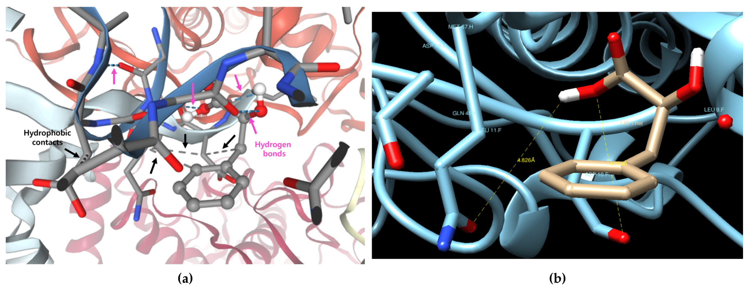
In the figure below: Predicting the binding form of phenyllactic acid (PLA) and mushroom tyrosinase (PDB ID: 2Y9X). PLA (shown in stick model, gray) is located within the catalytic pocket of tyrosinase. Dashed blue lines indicate predicted hydrogen bonds between PLA and the surrounding amino acid residues. Yellow highlights denote ionic interactions. (a) Visualization of predicted hydrogen bonds (blue dashed lines) and hydrophobic contacts (gray dashed lines) using the SwissDock server. (b) Measurement of key interactions at the top-ranked docking pose using the Chimera tool (Model 1).
