The stratum corneum (SC), the skin’s outermost layer, is vital for maintaining skin barrier function, offering protection against physical and chemical damage and preventing transepidermal water loss. This protective structure is maintained by a layered architecture, where specialized intercellular adhesion structures known as corneodesmosomes mediate the adhesion between corneocytes. The precise balance between the synthesis and degradation of these corneodesmosomes dictates skin desquamation.
A major challenge arises when this balance is disrupted, typically involving the excessive activity of serine proteases, specifically kallikrein-related peptidases (KLKs). Within the KLK family, KLK5 and KLK7 are known to degrade corneodesmosomal components (desmoglein 1, desmocollin 1, and corneodesmosin), thereby promoting SC desquamation. An abnormal increase in protease activity can lead to excessive degradation and impaired skin barrier function, which is implicated in inflammatory skin diseases such as atopic dermatitis and Netherton syndrome. In healthy skin, serine protease activity is tightly regulated by endogenous inhibitors like LEKTI (encoded by SPINK5), a major inhibitor of KLK5.
Shotokuseki extract (SE), a water-soluble substance derived from shale, is rich in various trace elements, including calcium (Ca), magnesium (Mg), aluminum (Al), iron (Fe), and zinc (Zn). While SE has been used in cosmetic formulations for over 40 years, its mechanisms of action, beyond promoting natural moisturizing factors and intercellular lipids, were previously unknown. Since several ions within SE, such as Ca and Mg, are known to be involved in epidermal differentiation and barrier formation, this study was established to clarify how SE influences SC homeostasis by regulating the synthesis of corneodesmosomes and the expression and activity of the proteases and inhibitors that control their degradation.
Methods
The study utilized a three-dimensional cultured human epidermis model for topical application of SE (1% or 5%) or a 5% Ion Mixture (IM), which contained the major biologically relevant ions found in SE at equivalent concentrations. Researchers analyzed the effects of treatment on gene expression (mRNA) for corneodesmosomal components, serine proteases, and protease inhibitors using reverse-transcription quantitative PCR. Protease activities (trypsin-like and chymotrypsin-like) were quantified using fluorogenic peptide substrates, and protein levels of corneodesmosomal components were assessed via Western blotting. Finally, histological analysis using H&E staining and Transmission Electron Microscopy (TEM) was performed to evaluate changes in epidermal morphology and structure.
Key Findings
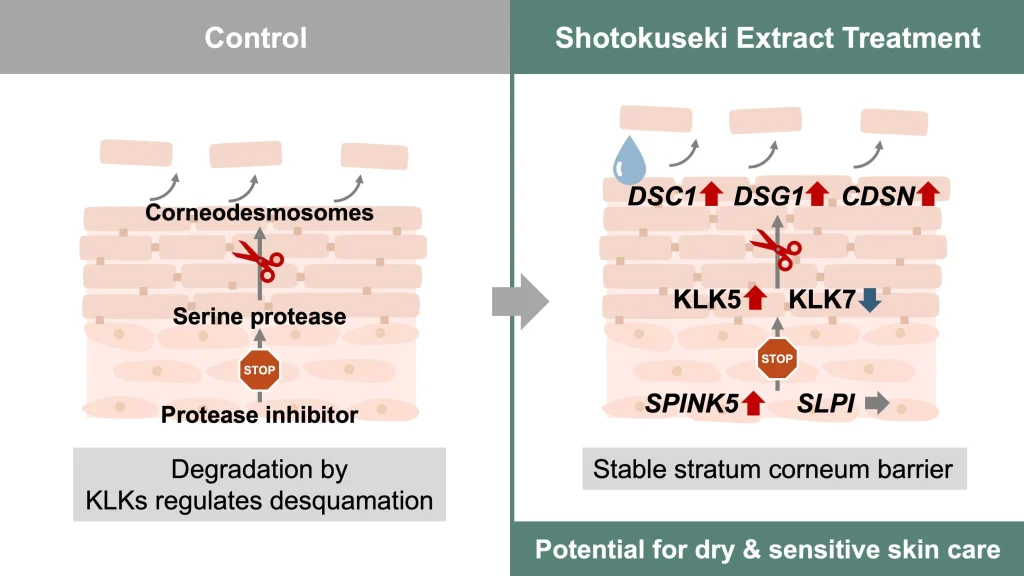
• Corneodesmosome Gene Upregulation: SE treatment (5%) significantly increased the mRNA expression of key corneodesmosomal components, including desmoglein 1 (DSG1), desmocollin 1 (DSC1), and corneodesmosin (CDSN).
• Protease/Inhibitor Gene Regulation: SE significantly increased the mRNA expression of both the trypsin-like protease KLK5 and its primary endogenous inhibitor, SPINK5 (encoding LEKTI).
• Selective Protease Suppression: SE significantly decreased the mRNA expression and activity of the chymotrypsin-like protease KLK7. Conversely, trypsin-like serine protease activity significantly increased with 5% SE.
• Chymotrypsin Activity Reduction by Ions: The decrease in chymotrypsin-like activity was observed not only with 5% SE but also with the 5% IM (Ion Mixture), suggesting that ions common to both solutions, such as calcium and zinc, may be involved in regulating KLK7 activity.
• Morphological Change: Histological analysis demonstrated that SE significantly increased the ratio of stratum corneum thickness to total epidermal thickness (SC/Total epidermis ratio), suggesting promotion of keratinocyte differentiation and SC structure formation.
• Protein Levels Unchanged: While gene expression was upregulated, no significant changes were detected in the protein levels of corneodesmosomal components (DSC1, DSG1, and CDSN) following SE treatment.
This research highlights the novelty of SE’s action as a multimineral preparation that orchestrates dual regulation within the SC. SE modulates key molecular pathways by simultaneously promoting the expression of genes encoding corneodesmosome components while regulating the protease–inhibitor system involved in their degradation. This effect is critical because the selective suppression of KLK7 activity, coupled with the increased expression of the KLK5 inhibitor SPINK5, is believed to effectively control the final stage of desquamation, thereby stabilizing the barrier function and improving the adhesive strength of the SC.
The findings provide compelling scientific evidence that SE can serve as an effective skincare ingredient for dry and sensitive skin conditions characterized by impaired barrier function, suggesting functional effects that extend beyond simple moisturization.
For future implications, the study recognizes several necessary steps. Although the results are highly encouraging, all experiments were conducted in vitro, necessitating in vivo validation to confirm the physiological relevance and barrier-modulating effects of SE. Furthermore, while SE promotes gene expression, the lack of significant changes in corresponding protein levels requires future studies to incorporate more frequent time points or complementary assays to understand the dynamic turnover and post-translational modifications of these proteins. Finally, comparative analysis with the IM suggested that trace elements specific to SE, beyond the major ions, might contribute to its effects, requiring further research to clarify the roles of individual ions within the complex mixture.
Link to the study: https://www.mdpi.com/1420-3049/30/23/4592