Exposure to ultraviolet (UV) radiation poses significant adverse effects on human health. UV light can be directly absorbed by cellular DNA, leading to the formation of dimeric photoproducts between adjacent pyrimidine bases, which can result in harmful outcomes such as mutagenesis, cell death, and skin cancer. The most common photolesions are cyclobutane pyrimidine dimers (CPDs), accounting for 80%–90% of the damage, and pyrimidine-pyrimidone (6-4) photoproducts ((6-4) PP), accounting for 10%–20%. While traditional sunscreens are important allies in avoiding the effects of UV radiation, they are a passive strategy that does not reverse existing photolesions; therefore, new active strategies to reduce UV-induced DNA damage are highly desirable.
The topical application of DNA repair enzymes, such as photolyase, represents an interesting and active strategy for skin photoprotection. Photolyases are flavoproteins capable of repairing CPDs and (6-4) PP damage by reversing the crosslinks using energy from blue light. However, the successful topical use of enzymes is limited because they are typically large and hydrophilic molecules that lack long-term stability and are sensitive to environmental conditions. Consequently, photolyase requires efficient delivery systems to penetrate the skin and exert its biological activities. Nanocarrier technology was considered a powerful tool to overcome these delivery drawbacks, enhancing stability in unfavorable environments, improving selectivity, and increasing tolerance to inhibitors.
Methods
This study investigated and compared three different nanostructured systems—polymersomes (PLs), liposomes (LPs), and polymeric nanoparticles (PNPs)—as potential carriers for the enzyme photolyase. Photolyase was produced recombinantly from Termus thermophilus in Escherichia coli and purified. PLs were produced by poloxamer 401 self-aggregation, LPs via the thin-film hydration method combined with extrusion, and PNPs using the double emulsion solvent evaporation method. Nanostructures were thoroughly characterized for size (hydrodynamic diameter, hD) and encapsulation efficiency (EE) using Design of Experiments (DoE) approaches, and subsequent in vitro studies evaluated enzyme release, cytotoxicity against human keratinocyte (HaCaT) cells, and the protective effect against UV irradiation.
Key Findings
• Encapsulation and Size: Both PLs and LPs showed favorable nanometric sizes (126 to 181 nm for PLs; 60 to 99 nm for LPs) and similar EE values (2% to 23% for PLs; 9% to 23% for LPs).
• PNP Performance: Polymeric nanoparticles (PNPs) achieved the highest EE, ranging from 85% to 96%. However, PNPs suffered from a slow enzyme release profile toward the external phase (approximately 15% after 10 days) and exhibited cytotoxicity against HaCaT cells, which were noted as the main disadvantages.
• Release Profile: Polymersomes (PLs) demonstrated the most adequate release, showing an initial burst release followed by slow, linear release, resulting in 37% release after 24 hours. This release percentage was twice that of LPs (17%) and three times that of PNPs (11%).
• Biocompatibility: LPs were found to be highly biocompatible, exhibiting 100% cell viability at concentrations up to 5 mg/mL, while PLs also showed low cytotoxicity, with concentrations below 12.5 μg/mL demonstrating no cytotoxic effects.
• UV Protection and DNA Repair: Cell viability recoveries following UV irradiation were significantly improved by nanoencapsulation, reaching 50%–60% viability with encapsulated photolyase compared to 28% without the enzyme.
• Superior Efficacy of PLs: Photolyase encapsulated in PLs demonstrated superior efficacy in protecting UV-irradiated cells, achieving a maximum of 50% cell viability (at 20 μg/mL) compared to 22% for LPs at the same concentration.
• CPD Repair: Nanoencapsulation in PLs significantly enhanced DNA repair, resulting in 81% repair of CPD lesions, which was double the repair percentage observed with the free photolyase (40%).
Conclusion
This study is groundbreaking as it proposes the first comparative evaluation of three innovative protein delivery technologies—PLs, LPs, and PNPs—for encapsulating pure photolyase. The research successfully demonstrated that nanostructures preserve or even increase the protective effect of the enzyme on UV-irradiated cells. Specifically, photolyase encapsulated into polymersomes (PLs) proved to be the most promising system, exhibiting superior efficacy in protecting cells from UV-induced photodamage, likely due to enhanced cell penetration and an adequate release profile. This research offers a promising and innovative pathway for the incorporation of photolyase into dermatological products, including sun care, skin care, and skin cancer prevention products. The successful optimization and comparative data lay a strong foundation for future translational efforts in creating active photoprotection strategies.
——————————————————————————–
To solidify the understanding of why encapsulation is necessary for photolyase: Think of the photolyase enzyme as a vital medicine carried across a hostile battlefield (the skin barrier and internal environment). Since the medicine (photolyase) is large and delicate, applying it directly risks it being destroyed or unable to cross the main fortification (the stratum corneum). The nanostructures (PLs, LPs, PNPs) act as tiny, armored transports. The Polymersomes, in this case, are the most effective transport: they are small enough to navigate the terrain, flexible enough to squeeze past obstacles, and are designed to release their payload efficiently where the damage (DNA repair) needs to occur.
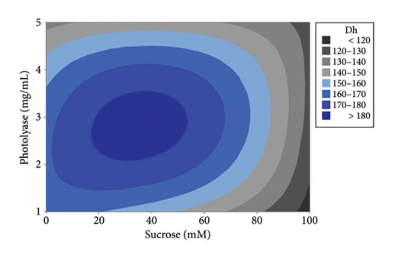
Contour plot, graph of the main effects, and Pareto chart for the (a–c) hydrodynamic diameter (hD) and (d–f) encapsulation efficiency (EE) based on the 32 experimental design applied to the preparation of photolyase-loaded polymersomes.
Link to the study: https://onlinelibrary.wiley.com/doi/full/10.1155/jnt/2317641