In the evolving landscape of cosmetic science, the limitations of traditional, surface-level observations—such as visual grading of skin texture or hydration—are increasingly evident. To achieve the rigor required for next-generation product development, we must pivot toward the cellular frontier. Microscopic morphological analysis serves as a critical solution, providing objective, high-fidelity data that traditional clinical photography cannot capture. By monitoring phenotypic transitions and cytoskeletal reorganization, we can identify measurable biomarkers of ingredient performance. These structural shifts, ranging from changes in individual cell geometry to alterations in colony architecture, offer definitive proof of cellular remodeling and extracellular matrix (ECM) modulation, allowing researchers to substantiate efficacy claims through the lens of fundamental cell biology.
Methodology
The laboratory protocols reflected in this study involve the cultivation of primary cell lines on standardized substrates to monitor real-time physiological responses to active compounds. Utilizing phase-contrast microscopy, we captured the distinct morphologies presented in panels (a) and (b), employing the 100μm scale bar as a primary tool for morphometric quantification. This imaging modality is essential for assessing cell-substrate integration, as it highlights the refractile properties of the cellular membrane. Adherence to these standardized protocols ensures that comparisons between treatment groups are based on quantifiable changes in confluency and spatial distribution.
Key Findings
A detailed comparative analysis of the phase-contrast micrographs reveals significant shifts in cellular behavior and mechanical engagement with the substrate:
- Cellular Shape and Phenotype: Panel (a) is dominated by rounded or cuboidal cell profiles. These cells exhibit a high degree of refractility at their edges, a classic indicator in phase-contrast imaging of low cell-substrate adhesion. Conversely, panel (b) demonstrates a profound phenotypic transition toward elongated, spindle-like morphologies, typical of activated fibroblast-like states.
- Cytoskeletal Stretching and Extensions: While the cells in panel (a) remain compact and constricted, panel (b) shows a marked increase in the development of bipolar and multipolar cytoplasmic extensions. These protrusive structures indicate a cell’s active “search” for contact and surface attachment. Notably, the 100μm scale bar confirms that the longitudinal stretching of these extensions in panel (b) spans distances significantly greater than the diameters of the clustered cells observed in panel (a).
- Distribution and Substrate Coverage: The spatial arrangement in panel (a) is characterized by “islands” or isolated colonies with high cell-to-cell adhesion but significant open space, suggesting a lack of migratory stimulus. In contrast, panel (b) displays increased substrate coverage and a more confluent, uniform distribution. This shift from clustered islands to an integrated network suggests that the treatment promoted cell migration and enhanced proliferative activity.
- Biological Implication: The transition from the refractile, clustered state in (a) to the spread-out, elongated state in (b) is indicative of optimized cell-substrate integration and a more active, migratory cellular state—primary benchmarks for validating “anti-aging” or “regenerative” cosmetic ingredients.
The ability to quantify these morphological shifts represents a paradigm shift in validating “scientifically-backed” skincare. By moving beyond anecdotal evidence toward cellular-level proof of efficacy, developers can confirm that active ingredients are driving meaningful biological processes such as fibroblast activation and migratory remodeling. This research approach allows the industry to transition from superficial claims to a model of precision dermatology. As high-resolution morphological data becomes the standard for efficacy testing, it will pave the way for personalized formulations that are tuned to the specific regenerative requirements of the cellular environment.
Link to the study: https://www.mdpi.com/2079-9284/13/3/126
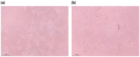
Microscopic images of HaCaT cells in (a) 1% DMSO and (b) 1 mg/mL perilla seed extract
after 24 h.