Cod liver oil is a potent source of omega-3 polyunsaturated fatty acids (PUFAs), such as EPA and DHA, which are essential for maintaining skin membrane fluidity and modulating inflammatory signaling pathways. However, the topical application of this oil is severely restricted by its extreme susceptibility to oxidative degradation, which leads to a loss of bioactivity, unpleasant odors, and potential skin irritation. Nanoemulsions were proposed as a potential solution because they can encapsulate sensitive hydrophobic compounds within nanoscale droplets, creating a protective microenvironment that mitigates oxidation. These systems also provide a high interfacial surface area that facilitates uniform contact and efficient interaction with the stratum corneum.
Methods
Researchers employed a 2^3 full factorial design to evaluate the combined influence of surfactant ratios (lecithin/PEG-15 hydroxystearate), processing methods (high-pressure homogenization vs. ultrasonication), and vitamin E acetate supplementation. Eight formulations were characterized for droplet size, polydispersity, ζ-potential, and encapsulation efficiency using dynamic and electrophoretic light scattering. Oxidative integrity was monitored through peroxide value determination and gas chromatography to analyze fatty acid preservation over a 60-day storage period. Furthermore, in vitro assessments included MTT-based cytotoxicity assays on human dermal fibroblasts and film-forming capacity tests using a modified occlusion model.
Key Findings
- Formulation F4, which utilized a 2.5:1 lecithin/PEG-15 hydroxystearate ratio and high-pressure homogenization (HPH), was identified as the optimal carrier for topical delivery.
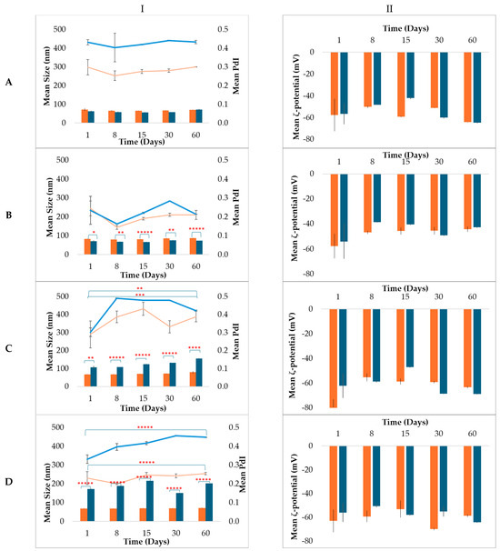
- The surfactant ratio was the dominant factor influencing stability, with higher lecithin content producing robust interfacial films that offered both electrostatic and steric stabilization.
- Efficiency of the emulsification process was formulation-dependent; HPH produced smaller droplets at higher lecithin ratios, while ultrasonication was more effective at lower ratios.
- The optimal nanoemulsion achieved a mean droplet size of 67.95 nm and a ζ-potential of −63.12 mV, maintaining colloidal stability across various temperatures for 60 days.
- Vitamin E supplementation significantly improved the oxidative resilience of the PUFA-rich lipid phase without negatively affecting the physical characteristics of the nanoemulsion.
- The formulations were found to be non-cytotoxic to human dermal fibroblasts, maintaining over 90% cell viability at all tested concentrations.
- The nanoemulsions exhibited a transient occlusive effect, with film-forming properties that provided an initial barrier but diminished over a 48-hour period.
The novelty of this research lies in its systematic demonstration that the performance of marine-lipid nanoemulsions is governed by the interplay between interfacial composition and processing conditions, rather than any single parameter alone. By establishing a rational framework for the stabilization of highly sensitive cod liver oil, the study overcomes a major barrier to the topical use of PUFA-rich marine lipids. Future implications of this work suggest a path toward specialized dermocosmetics for inflammatory skin disorders; however, subsequent research must include ex vivo skin permeation studies and in vivo evaluations of transepidermal water loss (TEWL) to confirm therapeutic performance.
Link to the study: https://www.mdpi.com/2079-9284/13/4/173
In the figure: Time-dependent evolution of cod liver oil nanoemulsions stabilized with lecithin/PEG-15 hydroxystearate at a 2.5:1 (w/w) ratio (■) or 1:1 (w/w) ratio (■) during storage at 25 °C for 60 days. Mean droplet size (bars) and polydispersity index (lines) are presented in column I and ζ-potential are presented in column II. Nanoemulsions were prepared by ultrasonication (panel (A): F1, F5; panel (B): F2, F6) and high-pressure homogenization (panel (C): F3, F7; panel (D): F4, F8), either without (A,C) or with (B,D) vitamin E. Data are presented as mean ± SD (n = 3). * p < 0.05, ** p < 0.005, *** p < 0.0005, **** p < 0.00005, ***** p < 0.000005.