The skin, being the body’s most extensive organ, faces constant degradation from internal factors like genetic predisposition and extrinsic stressors such as ultraviolet (UV) radiation and pollutants. A primary mechanism of skin aging is the accumulation of reactive oxygen species (ROS), which triggers inflammatory responses and activates mitogen-activated protein kinase (MAPK) pathways. This activation leads to the upregulation of matrix metalloproteinases (MMPs), enzymes that degrade structural proteins like collagen and elastin, resulting in wrinkles and loss of skin integrity. To address this, researchers explored a combination of Nicotinamide Mononucleotide (NMN) and Hyaluronic Acid (HA). NMN is known for its ability to boost intracellular NAD+ and regulate redox pathways, while HA is a well-established bioactive compound for extracellular matrix stability and moisture retention. The study hypothesized that their combined application would offer superior protective mechanisms against oxidative stress compared to individual treatments.
Methods
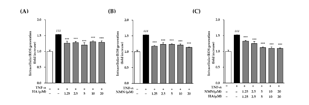
This study utilized human epidermal keratinocytes (HEKs) stimulated with TNF-α to induce an aging-related stress environment. Intracellular ROS levels were measured via DCFDA assays, while MMP-1 and Type I collagen (COL1A1) secretion were quantified using ELISA. Furthermore, Western blot analysis determined the phosphorylation levels of the MAPK subfamilies ERK, JNK, and p38. Finally, potential drug interactions were calculated using the SynergyFinder platform via Highest Single Agent (HSA) and Loewe additivity models.
Key Findings
- Enhanced ROS Suppression: Co-treatment with NMN and HA more effectively reduced ROS levels in TNF-α-stimulated cells than single treatments, particularly at lower concentrations.
- Inhibition of Matrix Degradation: The combination significantly attenuated the secretion of MMP-1, the chief enzyme responsible for collagen breakdown.
- Variable Collagen Stimulation: While HA increased COL1A1 levels at high concentrations, the combined effect was found to be concentration-dependent rather than uniformly synergistic.
- MAPK Pathway Regulation: Combined treatment modulated the phosphorylation of ERK, JNK, and p38 signaling, which are critical pathways that drive skin aging and inflammation.
- Concentration-Specific Synergy: Synergy analysis identified specific ratios, such as 2.5 µM HA and 5 µM NMN, as providing the highest predicted synergy for ROS inhibition.
The novelty of this research lies in its clarification of the complementary interactions between NMN and HA, demonstrating that they work through distinct but converging mechanisms—NMN through intracellular redox regulation and HA through receptor-dependent signaling. This suggests that their combination is a potent strategy for modulating oxidative stress and skin-aging responses. Future implications of this work include the development of more sophisticated anti-aging cosmetic formulations, though further validation is required using 3D co-culture models and in vivo animal studies to confirm these biological effects in more complex skin environments.
Link to the study: https://www.mdpi.com/2079-9284/13/3/116

In the figure: Hyaluronic acid (HA) and nicotinamide mononucleotide (NMN) mitigate the elevation of intracellular ROS triggered by TNF-α exposure. Human epidermal keratinocytes (HEKs) (1 × 10⁴ cells/well) were seeded in black 96-well plates and starved in DMEM serum-free medium for 24 h. Then, after a 1 h pre-incubation with (A) HA treatment alone; (B) NMN treatment alone; (C) com-bined treatment with HA and NMN. , cells were treated with TNF-α (20 ng/mL) and DCFDA (10 μM) cotreatment for 15 min to trigger ROS generation. Fluorescence intensity was then measured at 485/535 nm, with the control group receiving only DCFDA. The data are shown as means ± SEM of triplicate wells (n = 3). For all statistical evaluations, a threshold of p < 0.001 was established for significance; specifically, ### denotes a substantial difference relative to the untreated control, while symbols *** represent significant changes compared to the TNF-α-treated group at p < 0.001, respec-tively. White, black, and gray bars represent the control, TNF-α–treated, and compound-treated groups, respectively.