

Rising global temperatures and frequent heat waves driven by climate change present a growing threat to human skin health. Acute hyperthermia disrupts skin homeostasis, triggers inflammatory skin conditions like atopic dermatitis, and impairs the skin’s natural regenerative and repair capabilities. Crucially, elevated temperatures accelerate “thermal aging” by promoting the production of matrix metalloproteinases-1 (MMP-1), enzymes that degrade collagen and elastic fibers, resulting in wrinkle formation and loss of skin elasticity. To combat these environmental stressors, natural cosmeceuticals—specifically antioxidant carotenoids—have garnered intense research interest. The microalga Haematococcus lacustris (formerly Haematococcus pluvialis) is the richest natural source of astaxanthin, a potent antioxidant and anti-inflammatory agent. While astaxanthin has demonstrated systemic thermoprotection in other cellular models, its protective properties had not been evaluated at the skin level prior to this study, making H. lacustris extract (HLE) an ideal candidate to address this critical gap.
Methods
The researchers extracted bioactive carotenoids from stressed H. lacustris microalgal cysts to evaluate their protective properties. Using human HaCaT keratinocytes, they tested HLE’s impact on cell viability, ROS formation, MMP-1 gene expression, and wound healing. In parallel, they utilized the nematode C. elegans as an in vivo model to analyze systemic antioxidant activity, locomotor thrashing, and touch responsiveness. All experimental datasets were statistically validated using one-way ANOVA, Student’s t-test, and post hoc analyses.
Key Findings
- Intracellular ROS Reduction: HLE significantly counteracted the intracellular formation of reactive oxygen species (ROS) in human keratinocytes induced by hyperthermia (44°C), hydrogen peroxide, and UVA radiation.
- Equivalent Natural Potency: At equal concentrations, HLE demonstrated comparable antioxidant efficacy to synthetic astaxanthin in mitigating thermal and chemical oxidative stress.
- Subcellular Bioavailability: Treatment with HLE significantly increased total antioxidant activity (TAA) in both the cell membrane and cytosol of keratinocytes, showing a predominant enrichment within the cytosol.
- Mitigation of Thermal Aging Markers: Under heat stress, HLE successfully prevented cell death, completely restored baseline levels of collagen-degrading MMP-1 gene expression, and significantly accelerated wound closure.
- Systemic in Vivo Thermotolerance: In C. elegans, dietary intake of HLE reduced heat-induced ROS levels, preserved muscle function and movement (thrashing), and rescued touch-provoked responsiveness impaired by UVA stress.
This research establishes the novelty of HLE as a powerful thermoprotective agent, marking the first study to validate the protective effects of astaxanthin-rich microalgae against heat stress specifically at the skin level. By demonstrating that HLE can prevent cellular damage, suppress collagen-degrading enzymes, and promote wound healing, the study positions HLE as a highly promising cosmetic ingredient for mitigating climate-induced thermal aging. Future implications of this research highlight the need for further studies combining hyperthermia and UVA exposure to assess their cumulative or interactive effects on skin. Ultimately, these positive in vitro and in vivo results support the transition to testing HLE in three-dimensional reconstructed human epidermis models to confirm its real-world translational relevance.
Link to the study: https://www.mdpi.com/2079-9284/13/4/214
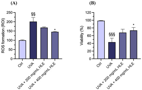
In the figure: Effects of HLE on ROS formation and touch-provoked responsiveness in C. elegans exposed to UVA irradiation. (A) N2 wild-type nematodes in the L1 stage were exposed to HLE 200 or 400 µg mL−1 for 48 h at 20 °C, then exposed to 20 J cm−2 UVA. After 24 h of recovery at 20 °C, ROS formation was assessed using the H2DCF-DA assay. ROS levels, indicated by fluorescence intensity, were quantified in the region of interest (ROI) in worms. (B) N2 wild-type nematodes in the L1 stage were exposed to HLE 200 or 400 µg mL−1 for 48 h at 20 °C, then exposed to 40 J cm−2 UVA. After 24 h of recovery, the touching assay was performed. Results are expressed as mean ± SEM (n. 30 nematodes). §§ p < 0.01 and §§§ p < 0.001 vs. standard condition; * p < 0.05 vs. nematodes exposed to UVA; one-way ANOVA, followed by Tukey’s post hoc test.