Why the outermost layer of skin is far from inactive
The Paradox at the Surface
The outermost layer of human skin is composed largely of dead cells.
At first glance, this seems biologically unremarkable—a layer of cellular remnants waiting to be shed.
Yet this same structure regulates water balance, controls molecular movement, organizes microbial interaction, and determines how the body interfaces with the external environment.
In skin biology, death does not mean inactivity.
It means specialization.
A Layer Designed to Stop Living
As keratinocytes migrate upward through the epidermis, they undergo a tightly regulated transformation.
Their nuclei degrade.
Organelles disappear.
Metabolic activity ceases.
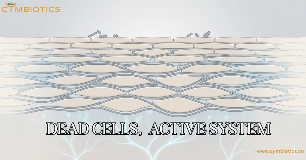
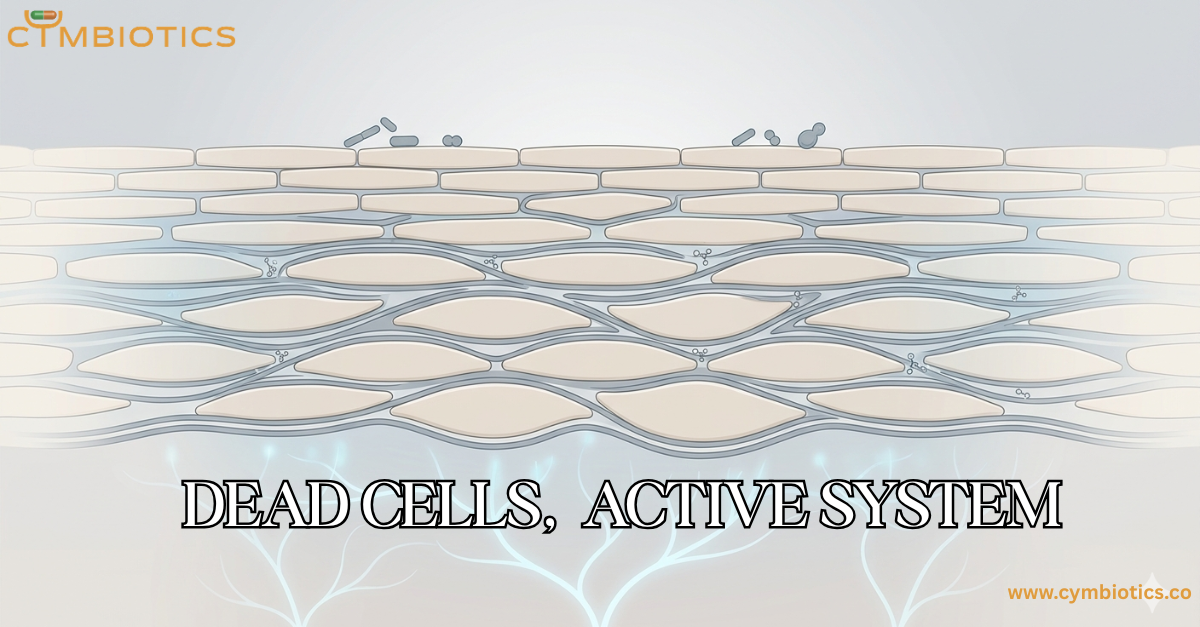
What remains are corneocytes: flattened, protein-dense cellular structures embedded within a highly organized lipid matrix.
This process is not degeneration.
It is engineering.
The skin intentionally creates cells that are no longer biologically active individually—but collectively form one of the body’s most functional interfaces.
Barrier Function Depends on the “Dead” Layer
The stratum corneum serves as the primary regulator of transepidermal water loss (TEWL).
Its architecture controls:
- hydration retention
- environmental permeability
- mechanical resilience
- diffusion of external molecules
This regulation depends not only on corneocytes themselves, but on the extracellular lipids surrounding them—primarily ceramides, cholesterol, and free fatty acids arranged in structured lamellar layers.
Together, they create a selectively permeable system.
Not fully sealed.
Not fully open.
Precisely organized.
Shedding Is a Controlled Biological Event
Even the loss of these cells is highly regulated.
Desquamation is governed through enzymatic activity involving proteases and corneodesmosomes, influenced by hydration gradients and local pH conditions.
The surface is therefore in constant transition:
- cells detach
- lipids reorganize
- barrier integrity recalibrates
What appears static is actually a continuous cycle of controlled renewal.
An Ecological Interface
The outer layer of skin also functions as a biochemical landscape for microbial life.
Surface lipids, hydration state, and corneocyte organization influence:
- microbial distribution
- nutrient availability
- local pH
- ecological balance
The microbiome does not interact with living epidermal cells first.
It interacts with the stratum corneum.
This means the so-called “dead” layer actively shapes one of the skin’s most dynamic biological ecosystems.
Implications for Topical Science
Every topical formulation first encounters this outer interface.
Not living tissue.
Not active vasculature.
But a highly ordered layer of terminally differentiated cells and extracellular lipids.
This makes barrier interaction fundamentally important to dermal delivery.
Effective delivery systems must navigate:
- lipid organization
- hydration variability
- selective permeability
- surface diffusion dynamics
Technologies such as Cetosomes™, designed with lipid-compatible architecture, can integrate more effectively within this structured environment while minimizing barrier disruption.
Similarly, FADD™ (Fast Acting Dermal Delivery) supports efficient transport across the stratum corneum without relying on aggressive penetration mechanisms.
The objective is not to overpower the barrier.
It is to work intelligently with one of biology’s most specialized surfaces.
The Most Active Layer May Be the Least Alive
The stratum corneum contains cells that no longer divide, synthesize proteins, or perform traditional metabolic functions.
Yet together, these cells regulate some of the skin’s most critical processes.
Hydration.
Protection.
Diffusion.
Microbial interaction.
Environmental adaptation.
The outermost layer of skin may be composed of dead cells.
But collectively, they form one of the body’s most active systems.
References (Verified & Correct)
- “Structure and Function of the Stratum Corneum Extracellular Matrix” – Peter M. Elias. Journal of Investigative Dermatology, 2012.
- “The stratum corneum comprises three layers with distinct metal-ion barrier properties” – Akiharu Kubo et al. Scientific Reports, 2013.
- “Presence of epidermal-derived thymocyte activating factor/interleukin 1 in normal human stratum corneum” – L. C. Gahring et al. Journal of Clinical Investigation, 1985.
- “Antibiotic Delivery Strategies to Treat Skin Infections When Innate Antimicrobial Defense Fails” – Ryan F. Donnelly et al. Pharmaceutics, 2020.