When oxygen is discussed in skin science, the conversation often stays broad. Skin needs oxygen for metabolism, repair requires oxygen supply, and circulation supports tissue vitality.
But skin does not experience oxygen uniformly.
Different skin compartments exist within distinct oxygen environments, creating physiological gradients that quietly influence metabolism, barrier behavior, repair pathways, and cellular signaling.
Not every skin cell breathes equally.
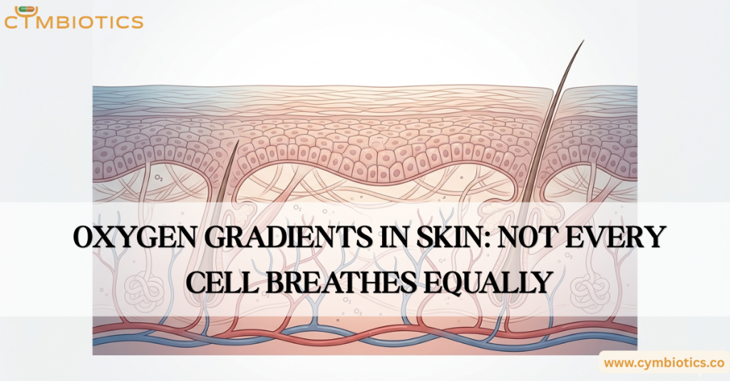
Skin possesses a unique oxygen arrangement. Unlike many tissues that rely almost entirely on vascular delivery, skin receives oxygen from two directions. Blood vessels within the dermis deliver oxygen from below, while atmospheric oxygen contributes at the surface. This creates a depth-dependent oxygen gradient across the tissue.
The epidermis is largely avascular, meaning its cells do not sit directly beside blood vessels. Oxygen must diffuse through surrounding tissue environments to reach keratinocytes. As oxygen availability changes across layers, cellular physiology changes with it.
These gradients influence metabolism.
Cells adapt their energy production strategies according to oxygen availability. Under higher oxygen conditions, oxidative metabolism efficiently generates ATP. Under lower oxygen conditions, cells may rely more heavily on glycolytic pathways and alternative metabolic adaptations.
In skin, this means cellular location partly shapes metabolic behavior.
Keratinocytes within different epidermal compartments can exhibit distinct metabolic states depending on their oxygen environment. Oxygen biology is therefore not simply a background condition. It helps determine how skin cells function.
Oxygen gradients also intersect with barrier formation.
The epidermal barrier depends on tightly regulated processes of keratinocyte proliferation, differentiation, lipid organization, and stratum corneum maturation. These processes occur within changing biochemical environments that include varying oxygen tensions.
Local oxygen conditions influence signaling pathways involved in epidermal differentiation and tissue organization. Barrier behavior is therefore shaped not only by structural proteins or lipid chemistry, but also by the surrounding metabolic environment.
The role of oxygen becomes particularly visible during repair.
In injured tissue, oxygen conditions can change dramatically. Reduced oxygen availability, or hypoxia, activates adaptive molecular pathways designed to help tissues respond to stress.
One major regulator is Hypoxia-Inducible Factor-1 alpha (HIF-1α).
This signaling pathway influences genes involved in angiogenesis, vascular remodeling, metabolism, and wound adaptation. Activation of vascular endothelial growth factor (VEGF) signaling can stimulate new blood vessel formation, helping restore nutrient and oxygen supply to damaged tissue.
Interestingly, low oxygen is not always biologically detrimental.
Controlled hypoxic signaling participates in normal repair physiology. The relationship between oxygen and skin function is therefore nuanced: both excessive deprivation and balanced adaptation matter.
Oxygen biology also intersects with inflammatory regulation.
Immune cells respond to oxygen availability. Tissue oxygen conditions can influence inflammatory signaling, oxidative balance, macrophage behavior, and stress responses during injury or irritation.
Oxygen is not merely fuel.
It functions as a biological signal capable of shaping how tissues respond to challenge.
At Cymbiotics, skin is approached as a layered physiological environment governed by gradients rather than uniform conditions. Understanding skin requires understanding how oxygen, nutrients, metabolites, and signaling cues vary across tissue depth.
Oxygen gradients illustrate an important principle of skin biology: healthy function emerges from coordinated complexity.
Skin is not one environment.
It is a landscape of microenvironments, and oxygen helps define them.
References:
- Oxygen in acute and chronic wound healing – Schreml S, Szeimies RM, Prantl L, Karrer S, Landthaler M, Babilas P. British Journal of Dermatology, 2010.
- Epidermal sensing of oxygen is essential for systemic hypoxic response – Boutin AT, Weidemann A, Fu Z, et al. Cell, 2008.
- Loss of epidermal hypoxia-inducible factor-1α accelerates epidermal aging and affects re-epithelialization in human and mouse – Rezvani HR, Ali N, Serrano-Sanchez M, et al. Journal of Cell Science, 2011.
- 2D luminescence imaging of physiological wound oxygenation – Schreml S, Meier RJ, Wolfbeis OS, et al. Experimental Dermatology, 2011.
- Lactate and oxygen constitute a fundamental regulatory mechanism in wound healing – Trabold O, Wagner S, Wicke C, et al. Wound Repair and Regeneration, 2003.