
Your skin may look still.
Under the microscope, it isn’t.
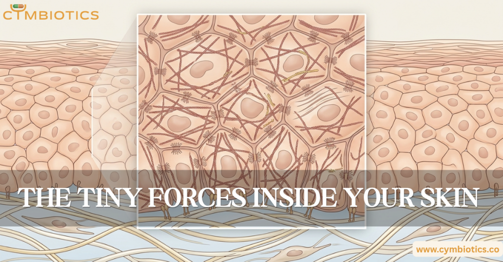
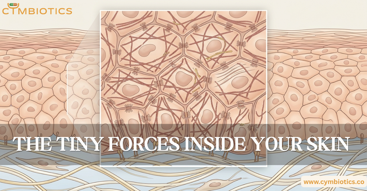
Every moment, individual cells are generating tiny mechanical forces. They pull on neighboring cells, tug against the extracellular matrix, change their shape, and sense the forces acting around them.
This microscopic tug-of-war helps determine how skin maintains its structure, repairs injuries, and responds to its environment.
Your Cells Have Their Own Machinery
Inside every cell is a dynamic protein network called the cytoskeleton.
It isn’t simply a framework that holds the cell’s shape.
The cytoskeleton constantly reorganizes itself and generates force.
One of its major components is actin, a filamentous protein that works together with myosin motor proteins.
Actin provides the tracks.
Myosin generates the pull.
Together, they create actomyosin contractility, allowing cells to produce mechanical tension within themselves and transmit forces to their surroundings.
Cells Pull on Their Neighbors
Skin cells aren’t isolated.
Keratinocytes are connected to neighboring cells through structures such as desmosomes and adherens junctions.
When actomyosin networks generate tension, some of that force can be transmitted across these cell-cell connections.
This means a mechanical change in one cell can influence its neighbors.
The epidermis therefore behaves less like a collection of individual units and more like a coordinated mechanical network.
The Matrix Pulls Back
Cells don’t only interact with one another.
They also attach to the extracellular matrix (ECM) through adhesion structures called focal adhesions and receptors known as integrins.
This creates another mechanical connection:
cell → adhesion complex → extracellular matrix
When a cell contracts, it can pull against the matrix.
The matrix, in turn, provides resistance.
That resistance becomes information.
When Force Becomes a Signal
This is where mechanotransduction comes in.
Cells can convert physical forces into biochemical signals.
A change in tension, stiffness, compression, or cell shape can influence intracellular signaling pathways.
One important mechanosensitive pathway involves YAP and TAZ, transcriptional regulators that respond to changes in cellular and tissue mechanics.
When the mechanical environment changes, these signals can influence processes such as:
- cell proliferation
- differentiation
- migration
- survival
- extracellular matrix production
Mechanical information can therefore become biological information.
The Skin Is a Mechanical Network
Imagine stretching a sheet of skin.
The force doesn’t simply affect the cells directly underneath your fingers.
Because cells are connected to each other and to the surrounding matrix, mechanical forces can travel through the tissue.
This interconnected architecture helps skin maintain its shape while allowing it to deform, recover, and adapt.
During wound healing, these forces become particularly important.
Cells migrate, contract, reorganize the extracellular matrix, and generate tension as the tissue closes and remodels.
The same machinery that maintains everyday tissue architecture becomes part of the repair process.
When the Balance Changes
Mechanical signaling is carefully regulated.
Too little force can alter tissue organization. Excessive or persistent mechanical stress can change cellular behavior and extracellular matrix remodeling.
This is one reason mechanobiology has become increasingly important in understanding fibrosis, scarring, aging, and tissue regeneration.
The physical environment isn’t merely something cells experience.
It can influence what they do.
The Cymbiotics Perspective
Skin biology isn’t governed exclusively by biochemical signals.
It is also shaped by physical forces.
Every keratinocyte, fibroblast, and connective-tissue cell exists within a mechanically active environment, constantly generating and sensing tension through its cytoskeleton, cell-cell junctions, and interactions with the extracellular matrix.
The result is a tissue capable of maintaining its architecture while continuously adapting to movement, injury, and changing mechanical conditions.
Beneath apparently still skin, millions of cells are constantly pulling, sensing, and responding.
References
The Roles of YAP/TAZ and the Hippo Pathway in Healthy and Diseased Skin – Rognoni E, Walko G. Cells, 2019.
Control of Cellular Responses to Mechanical Cues Through YAP/TAZ Regulation – Dasgupta I, McCollum D. Journal of Biological Chemistry, 2019.
Mechanobiology of YAP and TAZ in Physiology and Disease – Panciera T, Azzolin L, Cordenonsi M, Piccolo S. Nature Reviews Molecular Cell Biology, 2017.
Mechanoregulation and Pathology of YAP/TAZ via Hippo and Non-Hippo Mechanisms – Dobrokhotov O, Samsonov M, Sokabe M, Hirata H. Clinical and Translational Medicine, 2018.
Integrin-Mediated Mechanotransduction – Sun Z, Guo SS, Fässler R. Journal of Cell Biology, 2016.
Mechanotransduction at the Cell-Matrix Interface – Sun Z, Costell M, Fässler R. Trends in Cell Biology, 2019.