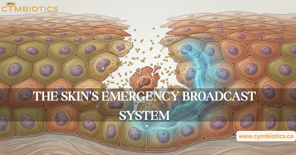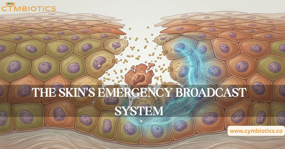

Damage one skin cell, and its neighbors can know about it within seconds.
You cut your skin.
The injury may be tiny, but the response isn’t.
Before new tissue is built, before immune cells arrive in large numbers, and before the wound visibly changes, cells around the injury begin communicating.
One of the earliest signals can be ATP, a molecule better known as the cell’s energy currency.
Outside an injured cell, however, ATP can become something else entirely:
an alarm signal.
When ATP Becomes a Signal
Healthy cells normally keep most of their ATP inside.
When a cell is damaged, ATP can be released into the extracellular environment. Nearby cells detect it using purinergic receptors, including receptors belonging to the P2X and P2Y families.
This allows extracellular ATP to act as a rapid chemical message.
The message is essentially:
Something has happened here.
This signaling can influence calcium levels, cellular movement, inflammatory responses, and other processes involved in early tissue repair.
The Calcium Wave
The message doesn’t necessarily stop with the first neighboring cell.
ATP released from an injured cell can stimulate nearby cells, triggering intracellular calcium signaling.
That signal can then propagate through the surrounding tissue as a calcium wave.
Think of it less as a single alarm and more as a rapidly spreading notification.
A local injury can therefore produce a coordinated response across cells that were never directly damaged.
Keratinocytes are particularly interesting in this process because they aren’t simply passive structural cells.
They can communicate with neighboring keratinocytes and participate actively in early wound signaling.
Purinergic Signaling: Cells Listening for ATP
The ability to detect extracellular ATP depends partly on purinergic signaling.
Purinergic receptors allow cells to respond to extracellular nucleotides such as ATP and ADP.
Different receptor types trigger different downstream responses.
Some open ion channels directly.
Others activate intracellular signaling pathways that can alter calcium concentrations and cellular behavior.
This gives skin cells a rapid communication system capable of converting tissue damage into biochemical information.
Not Just ATP
ATP isn’t the only molecule released when cells are damaged.
Injured or stressed cells can release a broader collection of molecules known as damage-associated molecular patterns, or DAMPs.
These include molecules such as extracellular ATP, HMGB1, and other intracellular components that become signals when they appear in the wrong biological context.
DAMPs help alert surrounding cells and the immune system that tissue damage has occurred.
Importantly, these signals don’t necessarily mean infection is present.
They are signals of cellular stress or damage.
Why the Message Matters
An injury creates a problem of coordination.
Cells need to know where the damage occurred.
They need to alter their behavior.
Some need to migrate.
Others need to proliferate.
Immune responses need to be coordinated.
The extracellular matrix needs to be remodeled.
All of this begins with communication.
Early ATP and calcium signaling can therefore help establish the local environment in which subsequent wound-healing events take place.
The first response isn’t necessarily about rebuilding the tissue.
It’s about telling the tissue that rebuilding needs to begin.
A Local Alarm With Tissue-Wide Consequences
The fascinating part is that the damaged cell doesn’t need to survive to communicate the problem.
Its surroundings can detect the molecular consequences of injury and relay that information.
This creates a chain of communication:
cell damage → ATP release → purinergic receptors → calcium signaling → neighboring-cell responses
Additional DAMPs and inflammatory mediators then contribute to the increasingly complex signaling environment.
What begins as damage to a microscopic area can therefore become a coordinated tissue response.
The Cymbiotics Perspective
Skin repair isn’t initiated by a single molecule or a single cell type.
It begins with communication between cells.
ATP, purinergic receptors, calcium signaling, DAMPs, and keratinocyte-derived signals form part of the early molecular language through which damaged skin communicates with its surroundings.
Long before a wound closes, the tissue has already started talking.
The first step in healing isn’t rebuilding the skin. It’s sending the message that something needs to be repaired.
References
- Mechanisms of Epithelial Wound Detection – Xu S, Chisholm AD. Cellular and Molecular Life Sciences, 2014.
- Calcium Signaling in the Photodamaged Skin: In Vivo Experiments and Mathematical Modeling – Rotenberg SA, et al. Scientific Reports, 2022.
- Ecto-Nucleoside Triphosphate Diphosphohydrolase 2 Modulates Local ATP-Induced Calcium Signaling in Human HaCaT Keratinocytes – Burnstock G, et al. PLoS ONE, 2013.
- Purinergic Signaling in Scarring – Burnstock G, Ulrich H. Purinergic Signalling, 2015.
- Alerting the Body to Tissue Injury: The Role of Alarmins and DAMPs in Cutaneous Wound Healing – Cordeiro JV, Jacinto A. Frontiers in Immunology, 2018.
- Air-Stimulated ATP Release from Keratinocytes Occurs through Connexin Hemichannels – Barr TP, Albrecht PJ, Hou Q, et al. PLoS ONE, 2013.