The epidermis functions as the body’s primary shield, regulating water loss and protecting against environmental stressors through complex programs of keratinocyte differentiation and transcriptional networks. A central regulator of this biology is the Vitamin D Receptor (VDR), which modulates the expression of essential barrier proteins such as filaggrin and loricrin, while also suppressing pro-inflammatory responses. However, the direct cosmetic application of active Vitamin D metabolites is severely limited by photoinstability, formulation challenges, and strict regulatory constraints. To overcome these hurdles, a synthetic VDR-activating peptide (VDR-Pep) was developed as a non-hormonal alternative. This solution was considered because peptides offer improved stability and formulation compatibility while specifically targeting the VDR ligand-binding domain to mimic the receptor’s beneficial physiological effects without the risks associated with steroidal compounds.
Methods
Researchers utilized HaCaT human keratinocyte cells to evaluate the peptide’s safety via CCK-8 viability assays and its efficacy through in situ proximity ligation assays (PLA) to visualize protein-protein interactions like VDR/RXR. Functional outcomes, including the expression of barrier-related proteins (filaggrin, involucrin, and loricrin) and the inflammatory marker IL-6, were quantified using Western blot analysis. Additionally, Fluo-4 AM staining was employed to measure intracellular calcium mobilization, a key driver of skin differentiation.
Key Findings
- Safety Profile: VDR-Pep demonstrated no cytotoxic effects at concentrations up to 200 ppm, making it suitable for cosmetic applications.
- Receptor Activation: The peptide effectively enhanced VDR/RXR heterodimerization, confirming the activation of the canonical signaling pathway required for gene transcription.
- Barrier Reinforcement: Treatment led to a dose-dependent increase in S100A3 and key structural proteins, including filaggrin, involucrin, and loricrin, which are essential for maintaining skin integrity.
- Calcium Mobilization: VDR-Pep stimulated intracellular calcium levels at a potency comparable to or exceeding that of active 1,25-dihydroxyvitamin D3.
- Anti-inflammatory and Antioxidant Effects: Under UVB-induced stress, the peptide significantly suppressed IL-6 expression and increased NRF2-associated transcriptional engagement, suggesting enhanced resilience against oxidative damage.
The novelty of this research lies in identifying a stable, non-hormonal peptide capable of not only activating the VDR signaling axis but also engaging in functional crosstalk with the NRF2 antioxidant pathway. This distinguishes VDR-Pep from conventional Vitamin D derivatives by offering a more cosmetically adaptable approach to skin health. The future implications of this study are significant, as it provides a foundation for developing next-generation functional cosmetics aimed at restoring the skin barrier in conditions like atopic dermatitis and protecting against photoaging. Future research must now focus on validating these effects in in vivo human skin models and optimizing formulations for commercial use.
Link to the study: https://www.mdpi.com/2079-9284/13/3/150
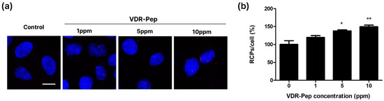
Enhancement of NRF2 transcriptional activation through increased NRF2/RNA polymerase II interaction in HaCaT keratinocytes. (a) HaCaT cells were treated with the VDR-activating peptide for 2 h. The interaction between NRF2 and RNA polymerase II (POL II) was evaluated using an in situ proximity ligation assay (PLA). Red fluorescent puncta (rolling circle products, RCPs) indicate individual NRF2/POL II complexes, and nuclei were counterstained with DAPI (blue). Magnification, ×600. Scale bars = 10 μm (b) PLA signals were quantified and expressed as rolling circle product per cell (RCP/cell) as a percentage of the control. Data are presented as mean ± SD (n = 3 independent experiments). Statistical analysis was performed using one-way ANOVA followed by Tukey’s post hoc test. * p < 0.05, ** p < 0.01 vs. control.