

Imagine cutting your finger.
Within seconds, thousands of biological events are set in motion. Blood begins to clot, immune cells arrive, and damaged tissue starts sending distress signals.
But perhaps the most remarkable event occurs over the following hours and days.
Millions of cells somehow know exactly where they need to go.
They migrate toward the wound, coordinate with neighboring cells, avoid healthy tissue, and collectively rebuild the damaged barrier. There is no central commander directing traffic, yet the process unfolds with extraordinary precision.
It may sound like science fiction, but it is one of the most fascinating realities of wound biology.
A Wound Creates a Biological Destination
When skin is injured, the wound immediately becomes a source of chemical signals.
Damaged cells release molecules known as damage-associated molecular patterns (DAMPs), while platelets and immune cells begin producing signaling proteins that spread through the surrounding tissue.
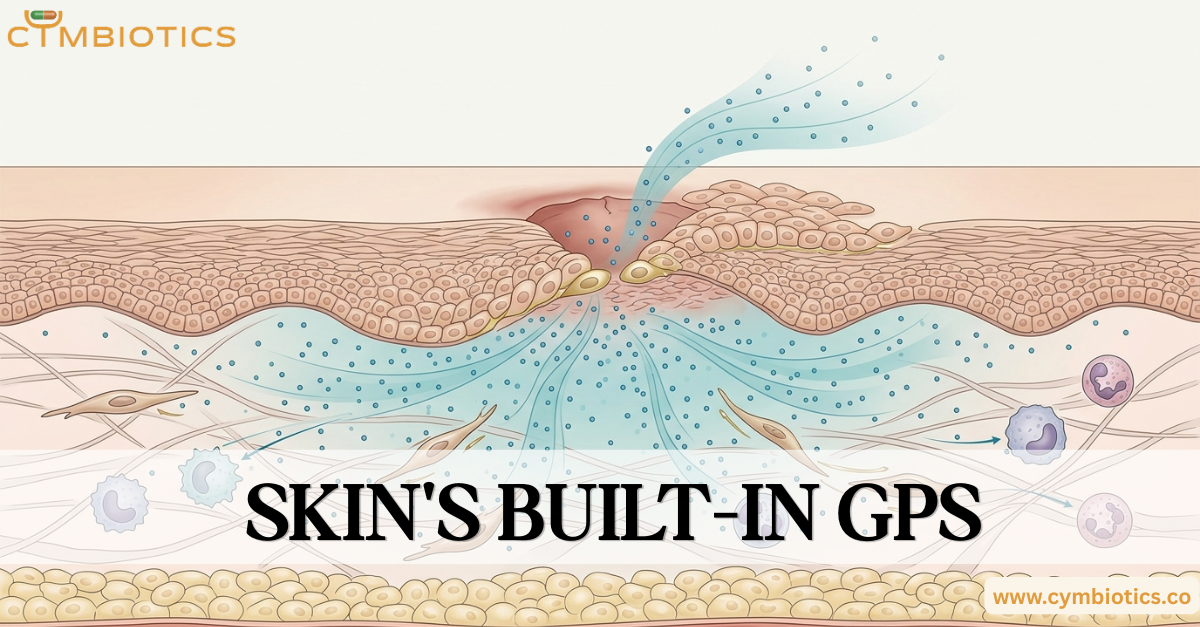
These molecules create concentration gradients around the injury.
The closer a cell gets to the wound, the stronger the signal becomes.
This process provides directional information that helps nearby cells identify where repair is needed.
Chemotaxis: Following the Signal
One of the most important mechanisms guiding repair is chemotaxis.
Chemotaxis is the ability of cells to detect and move toward increasing concentrations of specific chemical signals.
In wound healing, cells such as keratinocytes, fibroblasts, neutrophils, and macrophages constantly sample their surroundings through surface receptors. By comparing signal intensity across their cell membrane, they can determine which direction leads toward the injury.
In essence, cells are not randomly wandering.
They are navigating.
This chemical navigation system functions much like a biological GPS, allowing cells to locate damaged tissue with remarkable accuracy.
Growth Factors: The Navigation Beacons
Several signaling molecules help coordinate this migration.
Among the most important are:
- Epidermal Growth Factor (EGF)
- Transforming Growth Factor-β (TGF-β)
- Platelet-Derived Growth Factor (PDGF)
- Vascular Endothelial Growth Factor (VEGF)
These growth factors do more than stimulate repair.
They influence cell movement, proliferation, survival, and communication, helping create an environment that supports efficient tissue regeneration.
Different cell types respond to different combinations of signals, ensuring that the right cells arrive at the right time.
Re-Epithelialization: Rebuilding the Barrier
One of the most critical stages of healing is re-epithelialization.
This is the process by which keratinocytes migrate across the wound surface to restore the epidermal barrier.
To accomplish this, keratinocytes undergo temporary changes in behavior. They loosen some of their connections to neighboring cells, flatten their shape, and become more mobile.
The cells at the wound edge then begin moving across the injured area, forming a migrating sheet that gradually closes the defect.
Once the gap is sealed, these cells return to their normal differentiated state and rebuild the layered architecture of healthy epidermis.
Communication Prevents Chaos
If every cell simply moved toward the wound without coordination, repair would quickly become disorganized.
Instead, cells continuously communicate through chemical signals, adhesion molecules, and mechanical cues.
Fibroblasts help organize the extracellular matrix. Immune cells regulate inflammation. Endothelial cells form new blood vessels. Keratinocytes restore the barrier.
Each cell population responds to its own set of instructions while remaining integrated into the larger repair process.
The result is a highly coordinated biological response that restores tissue structure and function.
The Cymbiotics Perspective
At Cymbiotics, skin is viewed as a dynamic and highly intelligent biological system. Wound healing demonstrates that skin repair is not simply a matter of replacing damaged tissue. It is a carefully orchestrated process involving cellular communication, migration, and adaptation.
The concept of a built-in biological GPS highlights the sophistication of skin physiology. Through chemotaxis, growth factor signaling, and coordinated cell migration, millions of cells can locate a wound and contribute to repair with remarkable precision.
What appears to be a simple healing cut is, in reality, one of the most complex examples of cellular teamwork in the human body.
References
- Epithelialization in Wound Healing: A Comprehensive Review – Pastar I, Stojadinovic O, Yin NC, et al. Advances in Wound Care, 2014.
- Regulation of Wound Healing by Growth Factors and Cytokines – Werner S, Grose R. Physiological Reviews, 2003.
- Molecular Biology of Wound Healing – Velnar T, Bailey T, Smrkolj V. International Journal of Medical Research, 2009.
- Cellular and Molecular Processes in Wound Healing – Rodrigues M, Kosaric N, Bonham CA, Gurtner GC. Diseases, 2023.
- The Role of TGFβ Signaling in Wound Epithelialization – Ramirez H, Patel SB, Pastar I, et al. Advances in Wound Care, 2014.