
Millions of cells. Constant stretching. One remarkably cohesive tissue.
Think about everything your skin experiences in a single day.
You stretch, bend, scratch, sweat, exercise, sleep, and move. Your skin is constantly being pulled and compressed, yet the millions of cells making up its surface don’t simply separate.
So what holds them together?
The answer is a sophisticated system of cell adhesion structures that physically connect neighboring cells and allow the epidermis to behave as one mechanically integrated tissue.
The Epidermis Is More Than a Stack of Cells
The epidermis is made primarily of keratinocytes, arranged in layers that progressively differentiate as they move toward the surface.
These cells aren’t just sitting beside one another.
They are connected through specialized molecular structures that allow them to withstand mechanical forces while maintaining tissue organization.
Two of the most important players are cadherins and desmosomes.
Cadherins: The Molecular Connectors
Cadherins are cell-adhesion proteins that help neighboring cells recognize and attach to one another.
In the epidermis, several cadherins contribute to maintaining tissue architecture and regulating how keratinocytes interact.
Their importance goes beyond simply “sticking cells together.”
Cadherins can also participate in signaling pathways that influence cell differentiation, proliferation, and tissue organization.
So the connection between two skin cells is simultaneously physical and biochemical.
Desmosomes: The Skin’s Reinforcement System
If cadherins help establish cellular connections, desmosomes provide mechanical reinforcement.
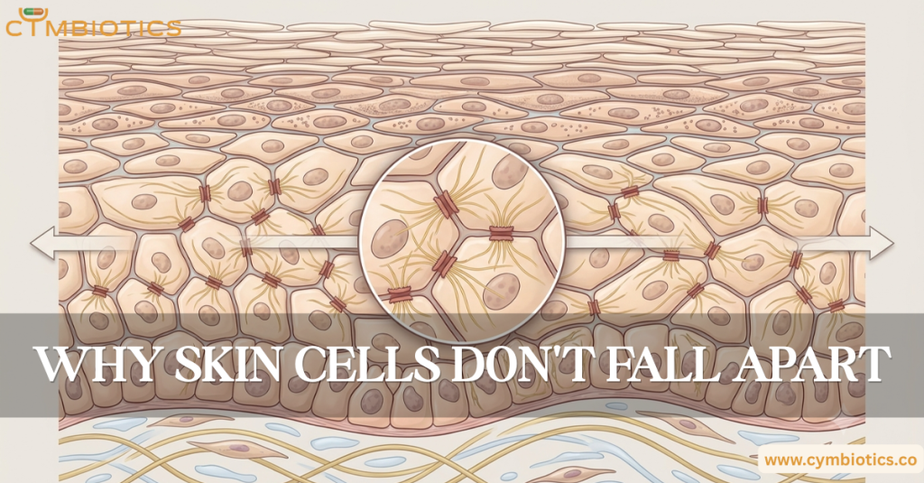
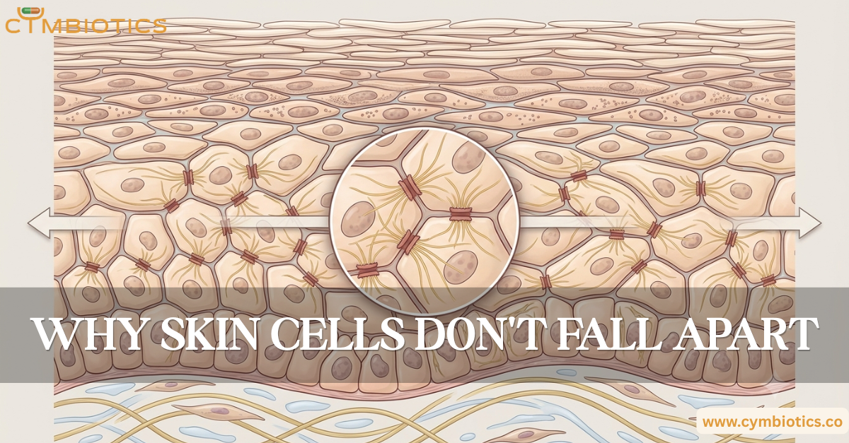
Desmosomes are specialized adhesion structures that connect the cytoskeletons of neighboring cells.
Inside a keratinocyte, keratin intermediate filaments attach to the desmosome. The desmosome then connects to the corresponding structure on the neighboring cell.
This creates a continuous mechanical network across the epidermis.
Instead of each cell absorbing mechanical stress independently, forces can be distributed across many connected cells.
That becomes particularly important in tissues such as skin that experience constant mechanical strain.
Tight Junctions: Sealing the Spaces
Not every cellular connection is primarily about mechanical strength.
Tight junctions help regulate the movement of substances between neighboring cells.
In the upper layers of viable epidermis, proteins including claudins and occludin contribute to these junctional structures.
They help control paracellular permeability, limiting uncontrolled movement through the spaces between cells.
Together with the lipid-rich stratum corneum, these junctions contribute to the skin’s barrier function.
When Mechanical Stress Arrives
Imagine stretching a sheet of skin.
The force doesn’t remain concentrated at a single point.
Because epidermal cells are mechanically interconnected, tension can be transmitted through cell-cell adhesion structures and the cytoskeleton.
This is where mechanobiology enters the picture.
Cells can sense mechanical forces and alter their behavior in response. Changes in tension can influence signaling, cytoskeletal organization, proliferation, differentiation, and tissue repair.
The epidermis therefore isn’t simply a passive protective covering.
It is a mechanically responsive tissue.
What Happens When Adhesion Fails?
When proteins involved in cell adhesion or desmosomal structure are disrupted, the consequences can be dramatic.
Cells can lose cohesion, making tissue more susceptible to mechanical separation and blister formation.
This is particularly evident in disorders involving autoantibodies against desmosomal proteins, where disruption of cell-cell adhesion can produce significant epidermal fragility.
The lesson is simple:
Skin strength depends not only on what its cells are made of, but also on how effectively those cells connect.
The Cymbiotics Perspective
At Cymbiotics, skin is understood as an integrated biological system rather than a collection of independent cells.
Cadherins, desmosomes, tight junctions, cytoskeletal networks, and extracellular matrix interactions allow millions of cells to function as a coordinated tissue.
The remarkable resilience of skin comes partly from this cellular architecture.
Every movement, stretch, and mechanical stress is handled by a tissue whose cells are physically connected, chemically communicating, and constantly adapting.
Your skin stays together because its cells don’t work alone.
References
- Desmosomes: Regulators of Cellular Signaling and Adhesion in Epidermal Health and Disease – Green KJ, Simpson CL. Cold Spring Harbor Perspectives in Medicine, 2014.
- Cell Adhesion in Epidermal Development and Barrier Formation – Rübsam M, Parameswaran H, Leight JL, et al. Current Topics in Developmental Biology, 2018.
- Epidermal Tight Junctions in Health and Disease – De Benedetto A, Kubo A, Beck LA. International Journal of Molecular Sciences, 2012.
- Structure, Function and Regulation of Desmosomes – Delva E, Tucker DK, Kowalczyk AP. Cold Spring Harbor Perspectives in Medicine, 2009.
- Mechanotransduction in Skin Inflammation – Chen Y, et al. Frontiers in Immunology, 2022.
- Keratinocyte Junctions and the Epidermal Barrier: How to Make a Skin-Tight Dress – Kirschner N, Brandner JM. Experimental Dermatology, 2012.