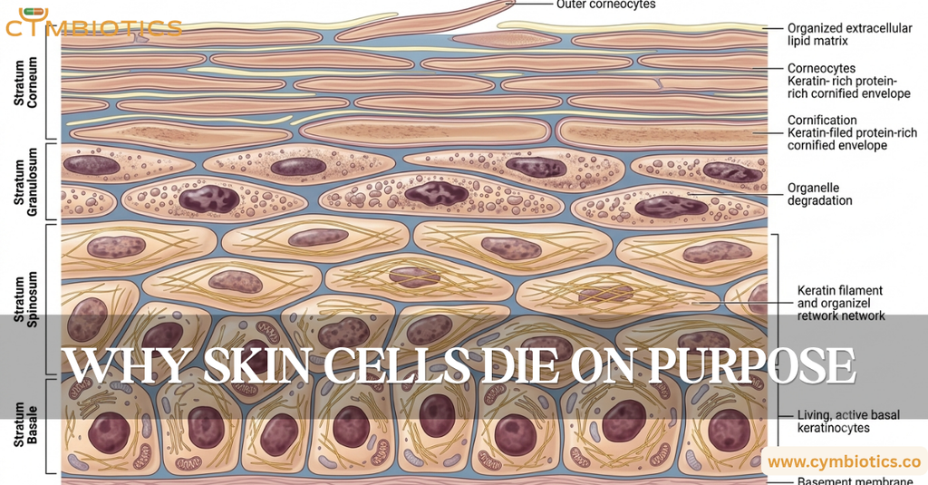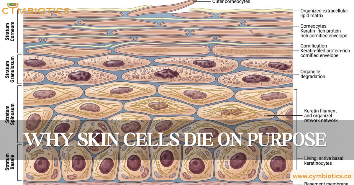

The controlled death of keratinocytes is essential for building a functional skin barrier.
Your skin is constantly making new cells.
But here’s the strange part:
Many of those cells are ultimately destined to lose their nuclei, organelles, and living cellular machinery.
And that’s not a failure.
It’s the plan.
As keratinocytes move upward through the epidermis, they undergo a highly specialized differentiation program called cornification. Their transformation into corneocytes creates the tough, protective outer layer that separates your body from the environment.
So in the epidermis, cell death isn’t simply an endpoint.
It is part of construction.
Not All Cell Death Is the Same
When we hear “programmed cell death,” we often think of apoptosis.
Apoptosis is a tightly regulated process in which a cell is dismantled in an orderly manner. Cellular components are fragmented, the cell is removed, and surrounding tissue is generally protected from uncontrolled damage.
Cornification is different.
Keratinocytes undergo a specialized terminal differentiation program in which they progressively transform into corneocytes, the flattened cells that make up the stratum corneum.
The destination is similar in one sense: the mature cell is no longer living.
But the biological process is fundamentally different.
The Journey of a Keratinocyte
The process begins in the basal layer of the epidermis.
Basal keratinocytes proliferate and produce daughter cells. Some remain within the proliferative compartment, while others begin a carefully regulated upward journey.
As they move through the epidermis, their biology changes.
They progressively alter their gene expression, cytoskeleton, organelles, and protein composition.
Keratin production increases.
The cells become progressively flattened.
Their internal architecture is reorganized.
Eventually, they enter the final stages of cornification.
A Cell Turns Into a Barrier
One of the most remarkable aspects of cornification is that the cell doesn’t simply disappear.
Instead, its remnants become part of the physical architecture of the stratum corneum.
During terminal differentiation, keratinocytes accumulate keratin filaments and develop a specialized protein-rich structure called the cornified envelope beneath the plasma membrane.
This envelope provides mechanical strength and helps create the structural framework of the outer epidermis.
Meanwhile, specialized organelles called lamellar bodies release lipids into the extracellular spaces between corneocytes.
These lipids help form the organized extracellular lipid matrix that is essential for limiting uncontrolled water loss and restricting penetration from the environment.
The result is a remarkable structure:
dead cells + organized proteins + extracellular lipids = functional barrier
Where Do the Organelles Go?
A mature corneocyte is very different from a living keratinocyte.
During cornification, cellular organelles including the nucleus and mitochondria are degraded through tightly regulated processes.
This is sometimes described as a form of programmed cell death, but it is more precise to describe cornification as a specialized terminal differentiation program with regulated cellular dismantling.
The distinction matters.
Cornification creates a specialized extracellular layer rather than simply eliminating unwanted cells.
The Final Product: Corneocytes
By the time keratinocytes reach the outermost epidermis, they have become corneocytes.
These flattened, protein-rich structures are densely packed within the stratum corneum.
They are embedded in an organized lipid matrix and connected through specialized structures called corneodesmosomes.
Eventually, these connections are enzymatically degraded, allowing individual corneocytes to detach from the surface through desquamation.
And the cycle begins again.
New keratinocytes are generated below while older corneocytes are continuously shed above.
Why This Matters
If keratinocyte differentiation or shedding becomes dysregulated, the architecture of the stratum corneum can change.
Cells may accumulate excessively, detach too quickly, or develop abnormal differentiation patterns.
This can contribute to disorders characterized by altered epidermal scaling, thickening, or barrier dysfunction.
The epidermis therefore depends on a carefully coordinated balance between:
proliferation → differentiation → cornification → desquamation
Too little or too much of any step can disrupt tissue homeostasis.
The Cymbiotics Perspective
Healthy skin isn’t maintained simply by producing new cells.
It depends on knowing when cells should proliferate, when they should differentiate, and how they should ultimately be transformed into the protective outer layer.
The apparent “death” of a keratinocyte is therefore part of a larger biological strategy.
Its final transformation creates a structure that protects the living tissue beneath it.
In skin biology, sometimes a cell has to stop being alive to become part of what keeps you alive.
References
- Cell Death in Skin Function, Inflammation, and Disease – Anderton H, Alqudah S. Biochemical Journal, 2022.
- Alternative Cell Death Mechanisms in Development and Beyond – Lippens S, Hoste E, Vandenabeele P, Agostinis P, Declercq W. Developmental Cell, 2011.
- Deconstructing the Skin: Cytoarchitectural Determinants of Epidermal Morphogenesis – Simpson CL, Patel DM, Green KJ. Nature Reviews Molecular Cell Biology, 2011.
- Pathophysiology of Keratinization – Schmuth M, Blunder S, Dubrac S, Elias PM, Gruber R. Journal of the European Academy of Dermatology and Venereology, 2018.
- Structure and Functions of Keratin Proteins in Simple, Stratified, Keratinized and Cornified Epithelia – Schweizer J, Bowden PE, Coulombe PA, et al. Journal of Cell Biology, 2006.
- Perspective: Controlling Epidermal Terminal Differentiation with Transcriptional Bursting and RNA Bodies – Wotherspoon DJ, Rogerson C, O’Shaughnessy RFL. Cells, 2020.